What's new
New features and improvements in version 2024
See the current Release Notes or all Release Notes for Neurolucida 360.
-
Update to offline software licensing. Due to updates to the Neurolucida 360 activation process, systems that that do not have access to the internet or have intermittent/unreliable internet access require reactivation. View instructions for offline software activation.
-
Improvements to the vessel tracing options in the 2D window.
-
Automove will now reposition focus when Continuous tracing is being used.
-
Color channel selection in puncta setup will begin at number 1.
-
Improvements to synchronization of contours delineated in 2D and 3D.
-
Image file paths are displayed in the Image Organizer.
-
During a movie, you can now change image-display settings for image slice and partial projections.
-
New ability to display the 3D scale bar while recording a Movie.
-
Other improvements and updates to the Movie mode interface have been implemented.
-
New Puncta-detection user interface improves and expands capabilities for specifying puncta detection settings.
-
Detect puncta based on proximity to other structures, such as trees, spines, and varicosities using rule sets.
-
Employ powerful And/Or criteria in your rule sets to find specific puncta easily.
-
Save your settings and reuse your puncta-detection criteria for multiple images, including in the batch pipeline for processing entire directories of image data.
-
-
Measure Intensity export will now report the centroid coordinate of any puncta.
-
You can now enable or disable the detection of spines and varicosities in the same location.
-
New ability to detect somas in the same location in different color channels.
-
The typical process width setting is now available in the automatic tracing setup to help optimize reconstruction when there is a large range in the size of branches and vessels.
-
New image filters added to the batch process pipeline.
-
ExM image filter includes three options to use to optimize modeling of your expansion-microscopy images.
-
Tone Mapping image filter uses contrast adjustments to reveal details in both bright and dim regions of your image.
-
-
The image display panel in the 3D Environment has an added option to eliminate the display of edge artifacts at the surface of 3D volumes.
-
Automatic stitching of overlapping image tiles in the Montage tool is now available to optimize the visual information of compiled image montages.
-
Improvements to the image support in the Measure Intensity tool.
-
Image support for the .czi format has been improved.
-
Slide scans from MBF Bioscience acquisition software can now be compiled.
-
Improvements to the movie creation tool:
-
You can now generate movies at high resolution for large data sets.
-
Use the image slice and partial projection views during movies to showcase important information.
-
Simple drag and drop to open.xml movie files.
-
SPARC Users
The Stimulating Peripheral Activity to Relieve Conditions (SPARC) program seeks to accelerate development of therapeutic devices that modulate electrical activity in nerves to improve organ function.
-
Term import and search has been added to tree and vessel classification for easier navigation of the anatomical terminology list
Previous Releases
-
The Neurolucida 360 Ultra package is now available! It includes all of the advanced vascular modeling features from Vesselucida 360 and enables you to explore the intricate dynamics of neuronal and vascular interactions with a single software package.
Learn more on our website or contact us for more information.
-
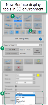
Surface effects of traced objects in the 3D environment now include enhanced options with the ability to include a legend, change colors, and adjust transparency for customized visualization and figures. This includes surface options for trees, vessels, spines, somas, puncta, and markers.
-
New contour-type legend can be displayed in the 3D window for enhanced visualization.
-
Existing puncta can now be excluded from subsequent detections with different detection parameters.
-
After applying image filters using the Batch Pipeline, you can now begin tracing right away.
-
Added the ability to save an image crop in .tif format increasing flexibility for use in other programs.
-
The Image Scaling dialog box that appears when opening images now has updated, more helpful information.
-
New Image Filters dropdown menu on the Image ribbon, includes filters for Correcting Illumination, Image Sharpening, and Background Subtraction.

-
To filter many images at once using your preferred filter settings, access the new Image Sharpening and Background Subtraction image filters in the Batch Pipeline. Once the filtering operation is complete, image files are automatically saved with the filename suffix of your choice.
-
Improved & expanded image support
-
Learn more from your 3D reconstructions using the new suite of surface-display tools, including by branch order, class and diameter. See the surface-display options
-
Use the Measure Intensity tool (on the Pipelines ribbon) to determine the average pixel intensity of modeled 3D structures or the image data inside a drawn contour for each color channel.
-
Use the Detect Orientation tool (on the Pipelines ribbon) to model the orientation vector of fibers in your images.
-
Faster image and data handling: Neurolucida 360 software uses a new approach to image and data handling based on intelligent image loading, maximized concurrent usage of CPU cores, multiple levels of data caching, and efficient use of RAM and GPU resources. Overall software function and performance is improved, highlights include:
-
Image- and data-files load noticeably faster than in previous releases
-
You'll notice that Neurolucida 360 software responds more quickly as you work; it handles hundreds of thousands of data points simultaneously.
-
Image adjustments are immediately displayed in both the 2D and 3D windows—without clicking any buttons.
-
-
New Batch Pipeline enables easy-to-use batch processing for image filtering and detection of neuronal structures
-
Simpler software authorization: Neurolucida 360 autofills the online Authorization Request Form with information about your computer system. Just fill in your name and contact information, plus the name of your PI to submit the form.
-
New subvolume tool in the 3D environment enables you to easily and systematically divide images/image stacks for analysis. It replaces the large volume reconstruction feature present in previous releases.
-
New Channel panel enables you to select one or more color channels to view, hide, and associate with structures in your images.
-
More options for selecting markers, contours, or vessels with the new Freehand Selection tools.
Previous Releases for SPARC users
-
SPARC vocabulary-term lists can now be displayed as ontological trees
-
New Import 3D Model tool for displaying generic SPARC organ scaffolds in the 3D environment
- Network licensing is now available for SPARC researchers.
- New Quick surface tool partially automates the process of creating contours to map interior and exterior surfaces.