Image Montage: How automatic montaging works
Tissue Mapper uses sophisticated algorithms that take features and overlapping into account.
Features
To match two images, the program uses features identified in the overlapping region of the two images. Features are points for which there are two dominant and different edge directions in a local neighborhood of the points. For a successful montage, the number of features identified must be relatively high.
In the figure below, montaging was successful because the algorithm found enough features (shown as green points) inside the overlapping region (the area between the dashed lines). The arrows show where the algorithm matched the points. 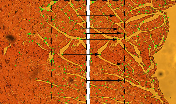
In the image below, however, automatic montaging failed because it found too few matching points within the region of overlap. Without these points, it couldn't complete the montage.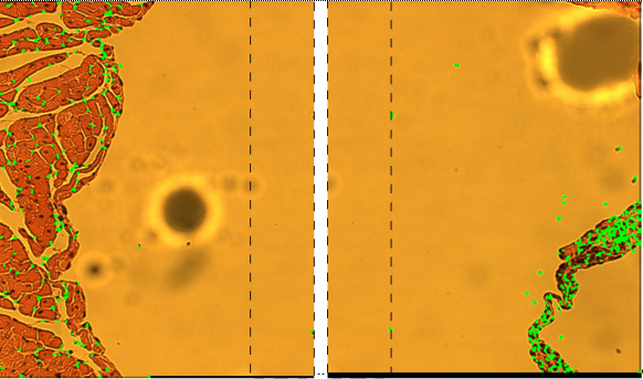
Overlapping
The next factor that affects automatic montaging is the amount of overlap (overlapping pixels) between images.
In general, a larger the overlapping region results in better performance and requires more computational time. We recommend an overlapping ratio of 10% to 25% of the image sizes as a starting point.
Sometimes, images are not included in the montage (they are "orphaned") because:
- The overlapping region is inadequate or the overlapping ratio is too large or too small.
- The images are unrelated to the other images used in the montage; you may have saved images in an incorrect location OR the images are from the same sample but have no related features.
- The images can't be automatically aligned in XY or Z because of an inherent property of the image.
Orphaned images are placed below the matched images. You can then align manually to override the automatic montage.
If you look closely, you'll see that the images don't align exactly. They almost look like they are tilting away from each other.
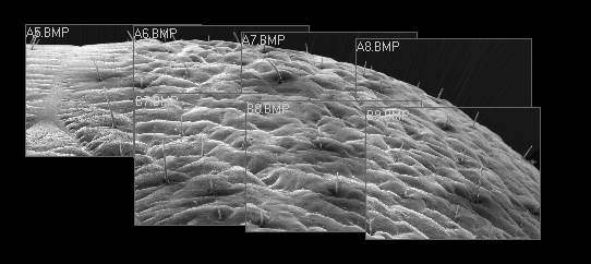
We can also see an incorrect Z alignment in the image below.
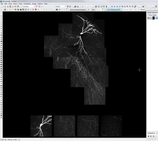
The software needs information from the X,Y alignment before it can perform the Z alignment. We allow alignment in X,Y, and also X,Y,Z, but the Z alignment happens internally after doing the X,Y alignment. It can't align just Z without knowing where it is in X,Y.

 +
+
