Isotropic virtual planes
Use this probe to estimate the ratio of total line length within an Optical Fractionator sampling scheme when isotropic or vertical sections are not practical.
Length can be determined for many different types of objects: tubules, nerve fibers, small blood vessels, microvilli, etc.
Try Spaceballs as perhaps a more robust alternative.
The probe is a set of parallel planes, and the counted parameter is the number of profiles that transect each plane within a defined counting region. The difference is that the planes are set at an isotropic angle to the tissue, emulating an IUR sectioning orientation.
Length measurements cannot be corrected for shrinkage, so the length reported is the final length in the processed tissue.
A series of sites are selected within each section by systematic random sampling. At each site, a 3D sampling box of constant volume is superimposed upon the slide to represent the boundaries of the counting area.
Although the sampling box is set up using Define Counting Frame, it is not technically a counting frame with rules of inclusion and exclusion as seen in other probes. The inclusion and exclusion zones are defined by the virtual planes.
A series of equidistant and parallel virtual planes with a random start are used to virtually section the 3D sampling box. The planes are represented in the program as a series of active lines (also called "moving lines"), that move across the section as the focal plane changes. The speed and direction of the active line movement across the screen depend on the angle of the virtual plane with respect to the X-Y plane.
- Thick sections: Sections need to be significantly thicker than the diameter of the fibers to be measured, and thick enough for several distinct optical planes through the section. Use Quick Measure Line to measure the diameter of the tubules if you think they might be thicker than the section thickness after processing.
- The structure of interest must be stained through the depth of the tissue section. Unstained tissue must be transparent enough to see the stained structure throughout the depth of the section.
- Thin focal planes, which are achieved using a high magnification, high numerical aperture lens, generally oil immersion.
- A contour traced around the region of interest.
Section thickness
- Use a high power objective lens with a high numerical aperture to focus at the top and bottom of a few sites throughout your sections to get a rough idea of the section thickness. This value can be edited later, but an approximate value is needed for setting up the 3D sampling boxes and guard zones.
- Click Probes>Tools>Define Counting Frame.
- Define a size that includes several profiles of the fiber(s) on which the length estimation is to be performed.
- If you are working with acquired images or image stacks, the counting frame needs to be smaller than the image in order to avoid the bias of an edge effect.
- Click Probes>Tools>Preview SRS Layout to preview the arrangement of sampling sites within the tissue section.
- Click in the tracing window to end the Preview mode.
- Start the Serial Section Manager and define the first section.
- At low magnification, draw a contour around the region of interest.
- Switch to an appropriate high magnification, then click Probes>All probes>Length>Isotropic Virtual Planes.
- If an SRS preview was used, the Scan Grid Size determined in the preview is automatically entered in the XY Placement of Counting Frames field.
- Isotropic Planes Settings: Enter a distance in microns between the isotropic planes. This distance is determined by the density and size of the profiles. This distance should be small enough that all profiles in the sampling box are intersected by at least one line. Also enter the desired number of orientations per sampling site. The distance between planes can be optimized during the pilot study.
- Distance from Section top to Optical Disector is the top guard zone height. This should be larger than the average thickness of the profiles to be counted and/or average tissue damage depth. It can also be set to a percentage of the measured section thickness, in which case you need to measure the section thickness at every sampling site.
- Sampling Box Height should be the minimal section thickness minus the top and bottom guard zones.
- Mounted Thickness is the actual z-depth of the sections as mounted on the slides (not the cut thickness). This should be measured with your microscope before starting. This value can be edited when viewing results if the initial estimate proves incorrect.
- Focus: Select manual focus.
- Select a marker to count transections.
- Focus through the section until all intersections have been counted.
- More than one plane orientation can be used to count each sampling box.
- To move to the next orientation, right-click and select Next Layout (which lists the layout number). The same sampling box is then resampled from a different orientation.
- Once all orientations have been completed, right-click and select Next Scan Site.
 Artificial Edge
If the section contains an artificial edge (a cut tissue edge or the edge of an acquired image), right-click and select Artificial Edge. The site is eliminated from the results calculation, and all markers in that site are disregarded by the software. The edge of the field of view in a live image is not an artificial edge, since the stage can be moved to see objects beyond the current field of view.
- Complete all sampling sites within the section.
- Move the stage to a new section, use the Serial Section Manager to designate a new section, and repeat the procedure.
Multiple orientations per site increases the efficiency of the probe by analyzing many different orientations within few sampling boxes rather than analyzing many sampling boxes, each with a new orientation.
When the probe is started, the software drives the stage to the first sampling site. The virtual planes are represented as lines that move as the focal plane is moved up and down. If the lines do not appear to move, that is because the virtual plane is orthogonal to the field of view; in essence you are looking down on the edge of the plane. When viewed within the sampling box depth, the active line is green within the sampling box, red on one side of the box, and blue on the other. Green indicates the inclusion line, red is the extension exclusion line, and blue is a 'line of indifference' used to indicate the position of the virtual plane outside the sampling box.
When the stage moves to the first counting box, the cursor appears as a diagram of a microscope objective with a slash through it and a + sign. This symbol indicates that the plane of focus is outside the counting frame as it lies in the sampling plane area. The active line is also completely red to indicate that the focal plane is above the 3D counting region. Focus down until the cursor becomes a simple crosshair again and begin counting.
View Overview Layout
At any time during the probe run, right-click and select Preview Isotropic Planes to see an overview of the probe layout. Stereo Investigator displays the full file with the sampling boxes overlaid on the tracing.
The current sampling box is indicated by flashing.
Click anywhere in the preview window to return to the current sampling site.

|
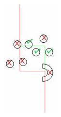
The "x" marker indicates a transection of a fiber by the green inclusion line. This represents one count. The red circle shows another transect, but is not counted because the fiber is more clearly in focus in the focal plane above, which happens to be at the top of the section in the guard zone. Therefore, that transect is not counted. The green circle shows that the fiber counted by the "x" is also transected by the red exclusion line in the same focal plane. However, since the fiber goes away from the line and comes back before being transected by the red line, the transect of the green line is still counted. |
- Only count intersections that lie within the bounds of the 3D sampling box (drawn in white).
- Each time a fiber cross section (referred to as a 'profile') is transected by the green active line (representing the isotropic virtual plane), a marker is placed on the point of intersection to count the profile.
- Do not count locations where the fiber merely touches the active line, the line must fully transect the fiber.
- For each profile intersection marked, follow the fiber through its full visible extent in current focal plane. If the fiber is continuously transected in the same focal plane by the active line until it runs into the exclusion zone (where the line is red), the original profile should not be counted.
- If the profile is transected by the green line, leaves the path of the active line, then is transected by the red exclusion line, the profile transected by the green line should be counted.
- If the fiber is transected by the green line in one focal plane, and then by the red line in another focal plane, it should still be counted -- unless the fiber intersection with the moving line stays in focus until the intersection moves to the red region of the line.
- If a fiber is transected multiple times by any one active line, but moves away from the line between focal planes, it should be counted at each intersection.
To help visualize this, remember that the fiber moving in and out of focus as it crosses the active line represents the fiber moving in and out of the sampling plane. If the sampling plane were the only visible part of the tissue, the fiber would appear to be segmented, and each fraction would be counted.
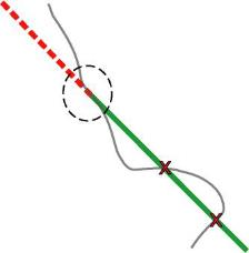
If the transect extends continuously from the green line to the red line in the same focal plane, then the transect is not counted, as illustrated here.

|
Here we see a sample fiber transecting the active line. The circled region shows the line continually transecting the active line at the boundary between the inclusion and exclusion regions. This transection is not counted. The two "X"s show two other complete transections of the line by the fiber, and both should be counted. Since the IVP is an estimator of length, not fiber number, the number of intersections per unit length is used. Thus, counting a single fiber multiple times is acceptable. A complicated part of the counting rule is found when the fiber is continually transected by the virtual plane from the inclusion region to the exclusion region. The difficulty is in recognizing this (uncommon) situation. The fiber will be in focus where it transects the active line in several subsequent focal planes. As the active line moves, the fiber stays in focus at the point of transection. The fiber is not necessarily in the same orientation as the active line, in fact, it can be in any orientation as long as each change in focal plane shows the fiber and moving line transecting in focus. If this is the case, and the fiber follows the active line into the exclusion (red) region, then the entire fiber is not counted. |
- Select Probes>Stereology results>Probe run list and click View Results.
Total Corners inside Region: Ccorners of the counting frames located within the contour of interest are used to calculate the total area for the Lv value.
Total Corners Possible: Number of counting frames multiplied by 4; total corners of all counting frames.
Average Area of Planes inside Boxes: Average area of the sampling planes contained within the counting frames.
Estimated Lv: Length/volume estimate result.
Estimated Total Length: Total length estimate for the entire volume sampled.
- Highlight the name of a section, then highlight a Run.
Layouts in Probe: Number of plane layouts used.
Corners in Probe: Number of counting frame corners (as used in the volume calculation) located within the contour of interest.
Total Possible Corners: Number of corners on all counting frames, whether they are located inside or outside the contour.
Fraction of Corners in Probe: Fraction of the total possible corners located within the contour of interest.
Total Plane Area in Probe: Total area of all of the sampling planes used.
Average Plane Area per Layout: Total plane area divided by the number of plane layouts.
Average Plane Area Per Sampling Box: Total plane area divided by the number of counting frames.