|
|
|
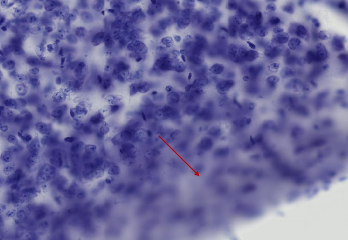
Problem 1:
Dark image  Poor lighting Poor lighting
|
Solution:
- Adjust light level and/or exposure level (see ).
- Acquire a new .
- Ensure that live image and background image are acquired at the same light level
|
|
Problem 2:
Bright specks consistently spaced throughout the
image
Background image contains
debris
|
Solution:
- Acquire another free of debris.
- Acquire a quick in the current field of view to ensure that the background image is suitable.
- Click the Acquire Image icon

|
|
|
|
|
Problem:
Over-saturated or “washed out" image
Light level is too high
|
Solution:
- Turn down the and perform the again.
- Ensure that the background image and live image are acquired at the same light level.
|
|
|
|
|
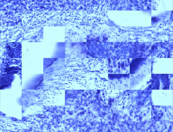
Problem:
Distinct horizontal and vertical seam lines at every image tile
No background image or background image with uneven lighting
|
Solution:
- Try enabling background correction (Acquisition>Enable Background Correction) or acquire another .
- Verify that lighting is uniform, especially around the edges of the image.
|
|
|
|
|
Problem:
“Doubling” or blurring along horizontal or vertical seam lines
Insufficient microscope calibration/lack of sufficient trim.
|
Solution:
- Verify that the microscope is properly calibrated (i.e., camera is aligned, lenses are grid-tuned, stage movements are functioning properly) - Virtual Tissue (slide scanning): Fine-tuning calibration
- Turn up the trim on the appropriate edges under the Virtual Tissue dialog box.
- If trimming does not work, a may be needed.
|
|
|
|
|
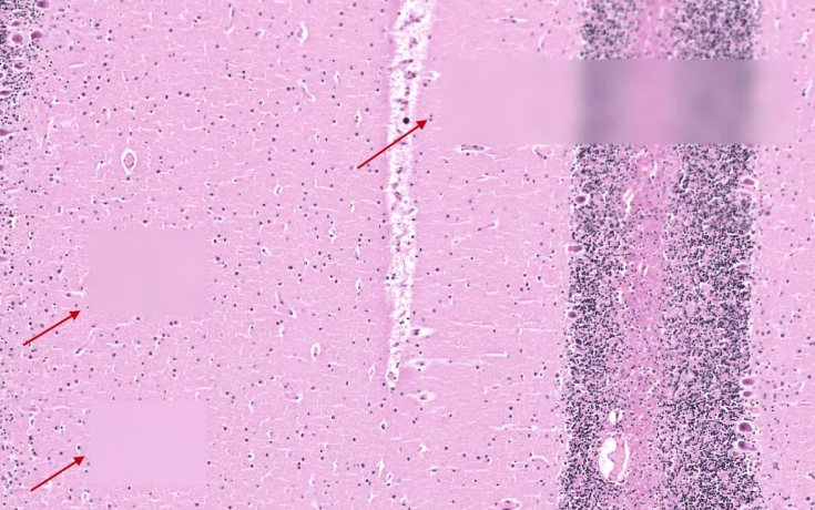
Problem:
Large out of focus areas in the image
Not enough focus sites
|
Solution:
- Try adding to the focus map.
- Verify that the focus sites are consistently dispersed throughout the contour or grid.
- If stage drift is the issue (and you allowed your microscope to warm up prior to acquisition), modify the settings by adding a manual focus interval. A manual focus interval enables you to adjust the focus during the imaging process, and to periodically update the focus map to compensate for drift.
- First, try adding a manual focus 3-4 times throughout the image and see if it helps (e.g., if the image has 2000 tiles, add a manual focus every 500 sites).
- Adjust as needed.
|
|
|
|
|
Problem:
Very distinct tile lines. Individual tiles are not lined up.
Mismatched objective and software lens
|
Solution:
- Verify that the lens selected in the software’s drop-down menu matches the objective selected on the microscope .
|
|
|
|
|
Problem:
Blatantly out of focus tiles or strips dispersed throughout an otherwise in-focus image.
In MBF’s experience, this occurs with extremely “wavy” tissue with drastic differences in z-depth between focus sites. The focus map interpolation cannot account for these large differences and out of focus tiles occur.
|
Solution:
- Try increasing the in the focus map to create a tighter triangulation grid.
|
|