Slide scanning: Troubleshooting for fluorescence microscopy
Uneven illumination / vignetting / tiling / lines
There are multiple possible reasons that could cause slide scan results to have distinct lines between the component image tiles, look unevenly illuminated or have vignetting effects, where each image tile has a . We recommend that you start by acquiring a 3x3 test image with all your channels, keeping the exposure times low (less than 200 ms to limit bleaching) and examine the results.
The issue is likely related to the light source. Here are some options to resolve this issue:
-
Make sure that your light source is centered and aligned. Because the procedure for this varies from light source to light source, we recommend that you contact your vendor, local microscope representative, or someone from the MBF Bioscience Tech Services team.
Many direct-coupled light sources (incl. Zeiss Colibri, X-Cite 120LED, X-Cite 120 Boost) do not have centering options.
Liquid light guide light sources (e.g., Lumencor SOLA) can potentially have problems with the liquid light guide cable. Most liquid light guide cable manufacturers recommend replacement every two years, regardless of usage. Replacements can be purchased through the manufacturer or MBF Bioscience.
-
Increase the Overlap percentage settings in step 5. Scan settings of the workflow. This is especially effective for mitigating vignetting effects.
Note that this may hide rather than fix the problem, will increase acquisition time, and may worsen bleaching of adjacent sites (see below). Typically an overlap of 10–25% is appropriate.
-
Enable Stitching XY in step 8. Sample acquire to preview the result and again in step 11. Start scanning for the actual slide scan operation.
-
Use background correction (step 7. Background correction of the workflow). This may alter the signal-to-noise ratio in your image.
-
Use the background subtraction filter (step 10. Setup filters of the workflow).
Bleaching of adjacent sites
Why bleaching occurs
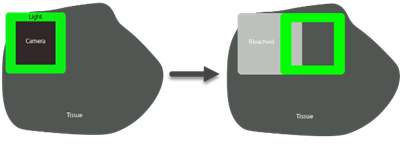
When scanning fluorescent tissue, the light source often covers a larger area of tissue than the camera’s field of view.
Any exposure of fluorescent dyes to light in the excitation range can cause bleaching; in slide scanning, “vignetting” or “lines” in the compiled slide scan image can result from "double" bleaching that occurs as images are captured from overlapping fields of view.
With fluorescence microscopy, it may be necessary to find an appropriate balance between adequate illumination, so that you can visualize structures that are labeled with the fluorescent dye, and minimal bleaching, to maximize the effective "lifespan" of the fluorescent dye used to label the specimen.
What you can do to prevent or mitigate bleaching
-
Improve tissue preparation
It may be worth investigating your tissue preparation protocol for opportunities to improve the stability of your fluorophores. If bleaching occurs in the DAPI channel (usually the most robust channel), there may be a problem with tissue preparation. Also keep in mind that some fluorophores are much more prone to bleaching than others.
-
Reduce the exposure time, then the illumination intensity
- In fluorescence microscopy, balance signal against bleaching
-
Decreasing the light-source intensity may help although LEDs typically run at 100% intensity to keep exposure times as low as possible.
Decreasing the light-source intensity will probably require a higher exposure time, which may result in a similar amount of bleaching, and will cause slower scans – meaning that there’s basically no advantage to lower light intensity in most cases.
-
Image Illumination Correction
 The Correct illumination tool (in the Image ribbon) may help to mitigate this issue.
 -
Aperture insertion into the light path (for Zeiss microscope systems)
Both apertures are shown in the photo for illustration only; they should NEVER be used together!
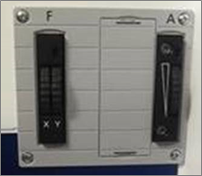
Zeiss makes two apertures: a square one (the XY) for the F slot, and a circular one for the A slot.
- If the F slot is not in use, insert the XY aperture in the F slot.
- If you have an ApoTome in the F slot, do not remove it; use the A slot instead to insert the circular A aperture.
- Only use the A slot for the aperture if you are equipped with an ApoTome.
- The A aperture may partially help with this issue, but it will not be as effective as the XY aperture.
- Closing down either of these apertures too much will exacerbate the uneven illumination.
Ruling out bleaching
To rule out bleaching as the cause for uneven illumination, we recommend acquiring an image with an Autofluorescent Plastic Slide (Chroma Technologies).
There are blank slides for each color channel. These slides provide uniform illumination and their intensity is even throughout the field of view.
The problem is with only one channel
-
Check to make sure that filter cube is properly seated and secure.
-
Other considerations:
- With high exposure times (above 300-500 ms), artifacts can appear, especially random red/green/blue pixels on color cameras.
- Older cubes do burn out, but we generally only recommend replacing a cube if, for example, there is no trace of uneven illumination for red and green, but blue shows uneven illumination, even with an appropriate exposure time and a known appropriate light source.
- Some light sources (such as the X-Cite 120 LED) use different bulbs for UV (DAPI) and other color channels, so it's possible (albeit rare) to have a light source issue that occurs only in one channel.
Try using the focus map's heat map (step 9. Define focus map) to look for large changes in Z before starting the slide scan. Large Z differences between sites can cause out of focus sites that look like uneven illumination.
Note that it is common to have some areas of the tissue that are brighter or darker than others. Optimize your camera histogram for the brightest area you are interested in, and adjust it post-acquisition using the Image Adjustment panel to improve the appearance of dimmer areas (typically using gamma adjustment).
Increase the number of focus sites only when areas are darker because they are out of focus, and not simply because they are dark.
Other issues
-
"Doubling" or poor XY alignment between sites: To solve this issue, review your calibration.
-
The image is too dark, too bright, or grainy: Adjust the histogram (this can be done before the acquisition [see Camera Histogram], or after the acquisition [see Image Adjustment ]). For best practices, refer to this tutorial video: Fluorescence imaging.
-
Out of focus areas: The is most likely the result of thermal drift. To avoid this issue, power your hardware on at least 2 hours before you set up your focus map and make sure that there is no hot or cold air blowing on it.
You may also need to add sites to your focus map. See step 9 of the slide scanning workflow.