Virtual Tissue (slide scanning): Optimizing your workspace for acquisition
The first step is to set up your workspace to have the tools you need ready for use. We recommend the following:
|
Tool |
Location |
Note |
|
AutoMove |
|
|
| Macro View |

|
Use to view contour tracings and navigate around the slide. |
| Image Organizer | Image>Image Organizer or
|
Use to navigate easily between the images or background images acquired. |
| Z-meter |

|
Useful when focusing through the tissue to generate 3D virtual slides. |
| Image Acquisition window | Acquisition>Acquire Image Stack | 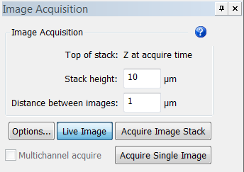
|
| Camera Settings | Acquisition>Video Tool
Panels>Camera Settings or  |
|
| Camera Histogram | Acquisition>Video Tool
Panels>Camera Histogram or
|
|
| Multichannel Control | Acquisition>Video Tool
Panels>Multichannel or |

![]() To optimize space: Dock the windows on the side of the screen (How do I set up my docking windows?) OR If a second monitor is available, move the Macro View and Video Tool Panels onto the second monitor to maximize the field of view on the primary monitor.
To optimize space: Dock the windows on the side of the screen (How do I set up my docking windows?) OR If a second monitor is available, move the Macro View and Video Tool Panels onto the second monitor to maximize the field of view on the primary monitor.