SLAP2 – Two Photon Microscope
A revolutionary 2-photon microscope for imaging neuronal activity at unprecedented speedProduct Overview
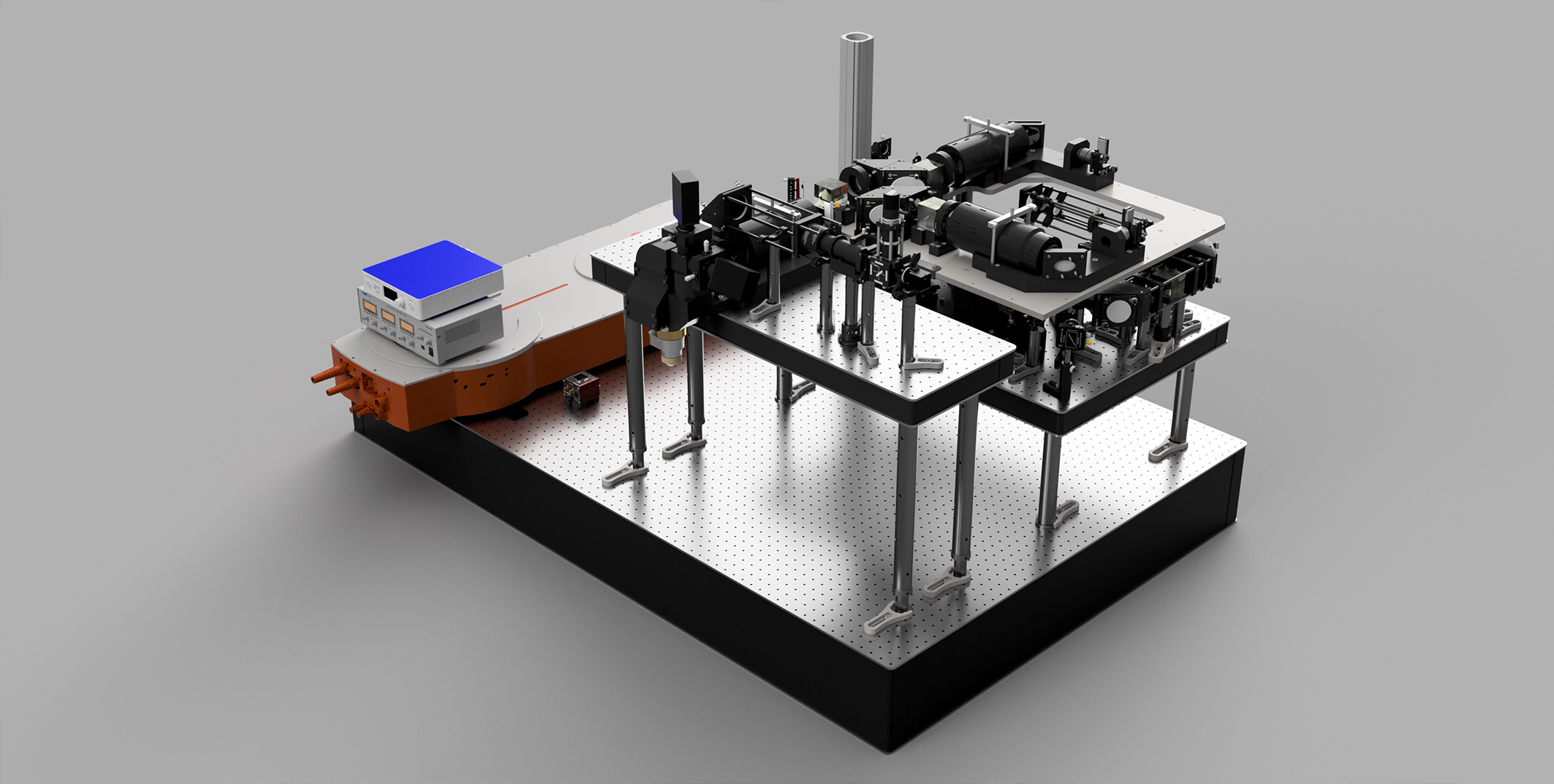
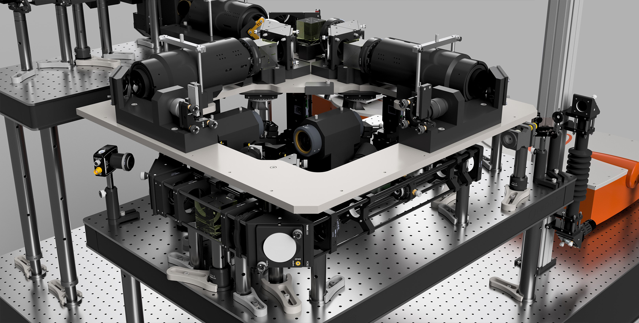
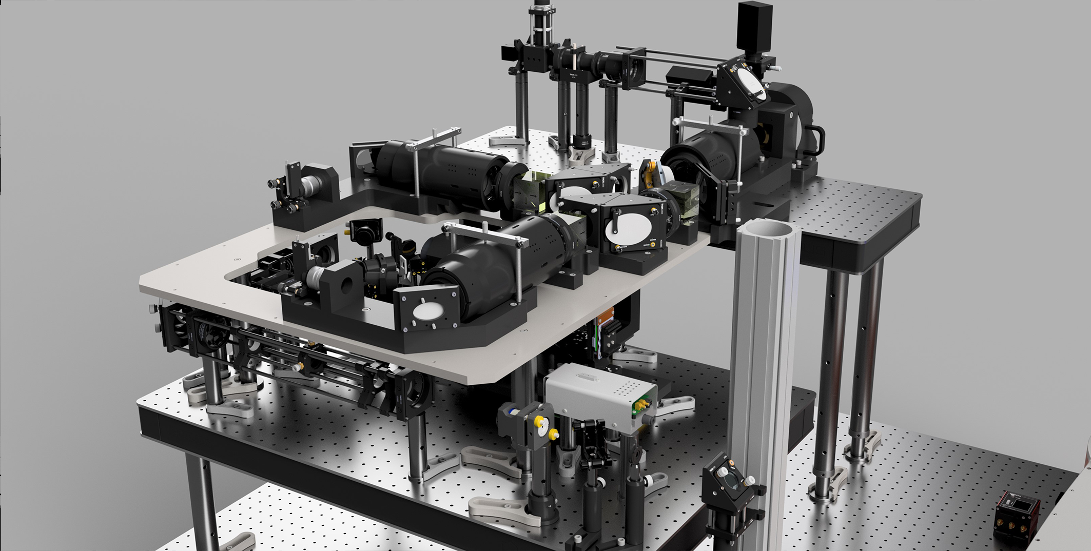
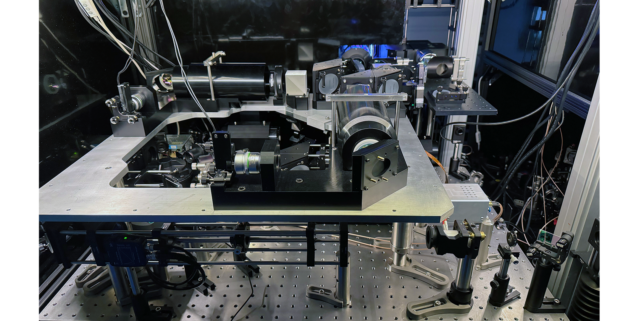
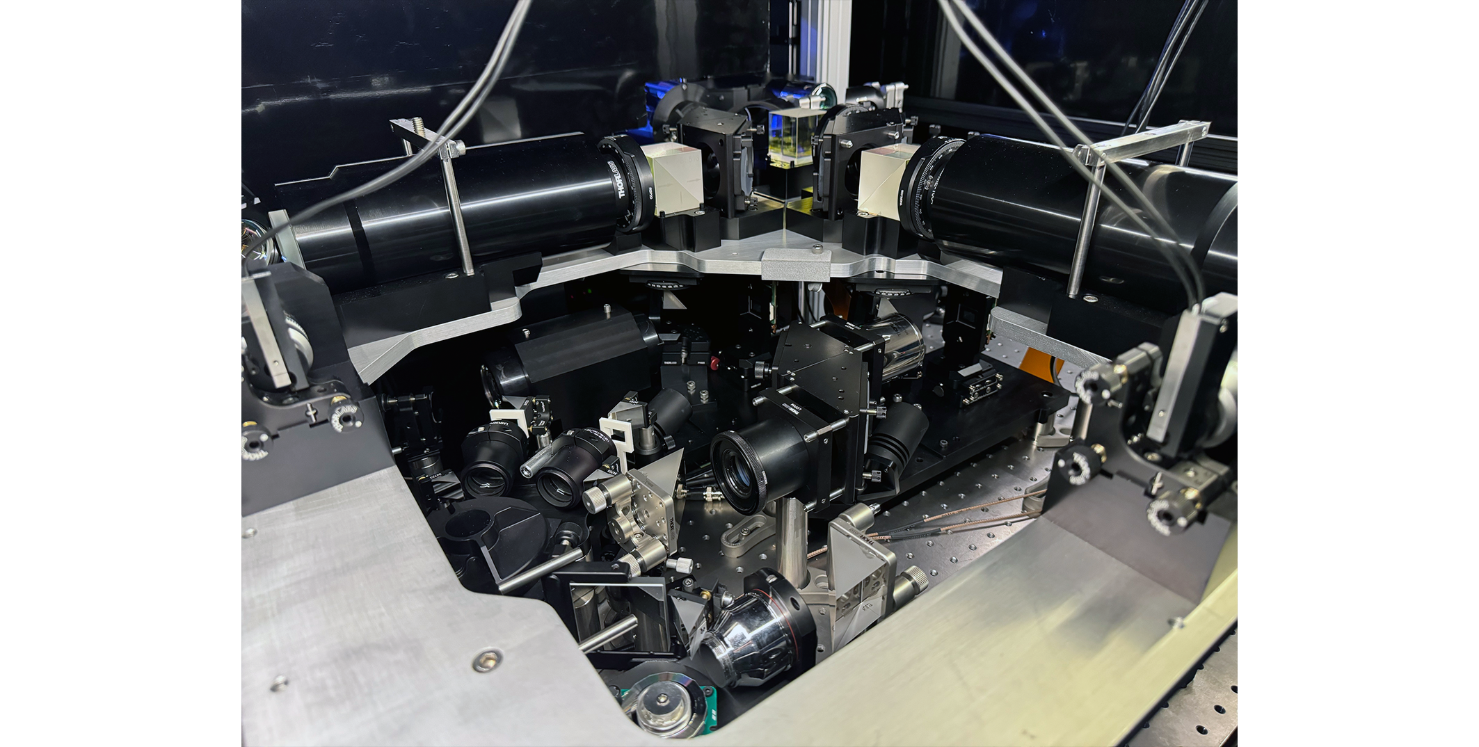
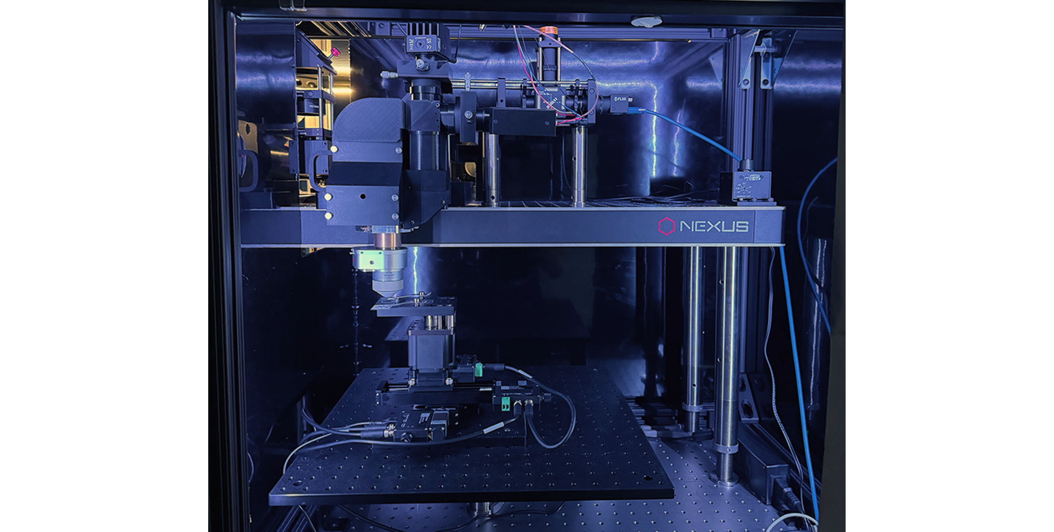
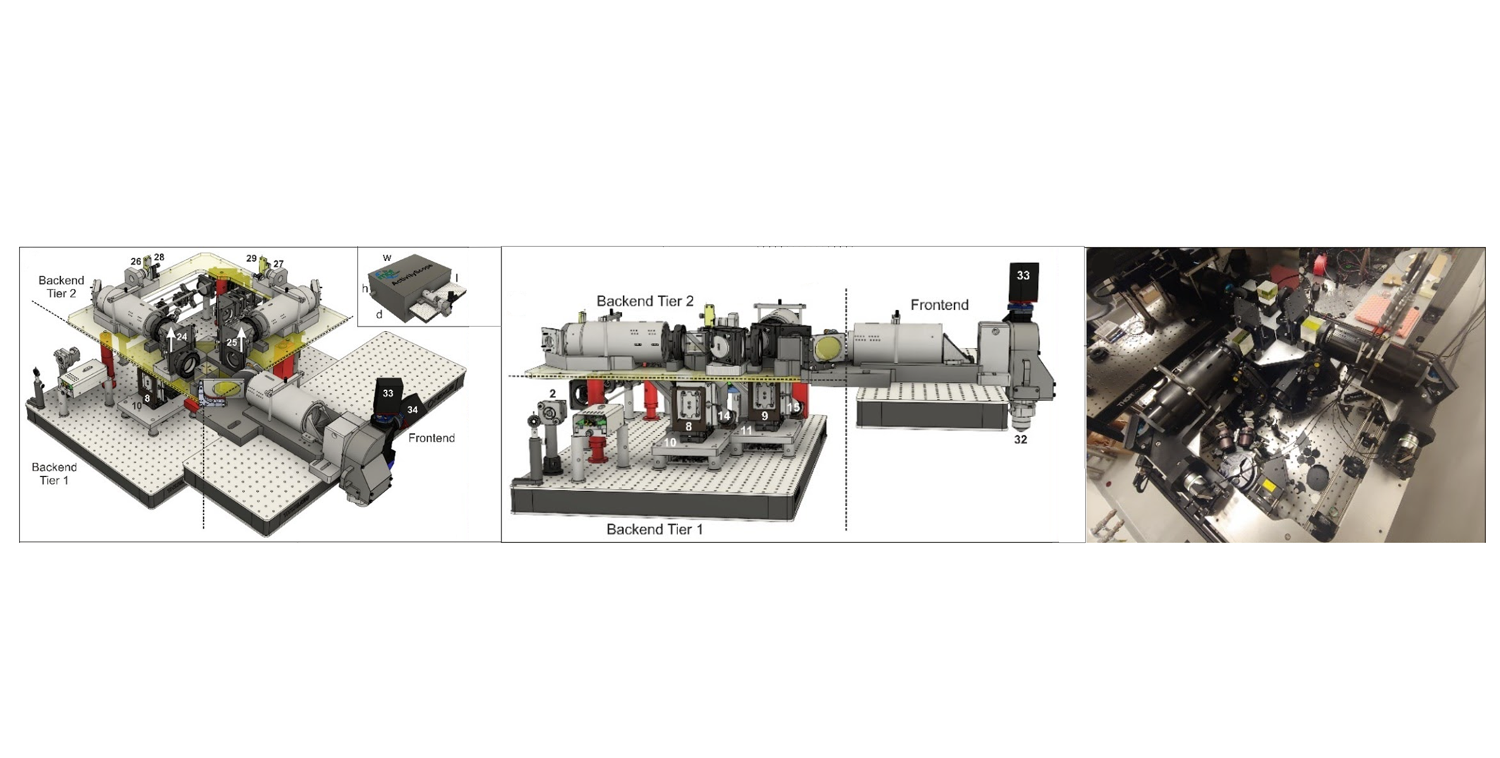
SLAP2 is a revolutionary new microscope for fast and flexible two-photon imaging, developed at the Allen Institute for Neural Dynamics and the Howard Hughes Medical Institute’s Janelia Research Campus. SLAP2 is optimized for cutting-edge in vivo synaptic imaging and population voltage imaging in three dimensions.
Key Benefits
- Perform optical imaging of neuronal activity at subcellular spatial resolution and temporal resolution in the millisecond range
- Sub millisecond temporal resolution (>10 kHz) for population voltage imaging in vivo and in vitro
- Image hundreds of synapses at >200 Hz with neurotransmitter indicators
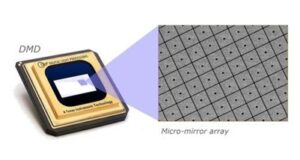
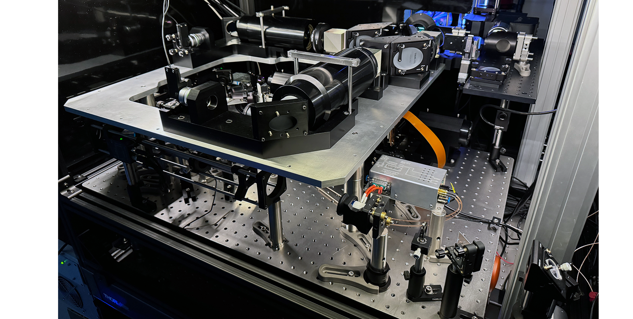
SLAP2 is based on a new laser scanning engine, called Random Access Projection Microscopy. The scan engine produces a vertical line of laser light and scans it horizontally across the surface of a digital micromirror device (DMD), illuminating one column of pixels at a time. Within each column, light hitting the ON pixels is relayed to the sample, while light hitting the OFF pixels is discarded.
All ON pixels in a given column are illuminated at once to produce a single measurement, so each line sweep is vertical projection of the sample at the ON pixels. After each sweep, the pattern on the DMD is updated, allowing a new projection to be collected on the next sweep. This programmable scan system makes SLAP2 faster and more flexible than previous two-photon microscopes. SLAP2 fills a unique niche for rapid, random access measurements in three dimensions, such as imaging synaptic activity in the dendritic arbors of individual neurons, or imaging membrane potential in neural networks.
| Excitation N.A. | Diffraction limited resolution at N.A. >0.9; Axial resolution <2.5 µm FWHM. |
| Collection N.A. | 1.0 |
| Field of view, lateral | 300 µm x 200 μm for each independently-targetable imaging path; 1200 x 800 pixels, 250 nm pixel pitch |
| Field of view, axial | >400 µm of aberration-free axial defocus via remote focusing. Independent remote focusing for each field of view. >100 Hz sine wave |
| Immersion | Water (SLAP2 is provided with a specific imaging objective not interchangeable with standard objectives) |
| Excitation Wavelength | 1030 nm (SLAP2 is designed to work with high-powered fiber lasers at this wavelength. We do not currently support other wavelengths) |
| Independently-steered fields of view (FOVs) | 2 |
| Frame rate | The frame rate in each column of each field of view is the line rate (10.8 kHz per field of view) divided by the number of pixels in that column. Each integration ROI counts as only one pixel. |
Other information: See US Patent application US20220382031A1
Download SLAP2 product sheet here.
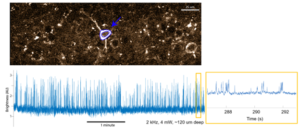
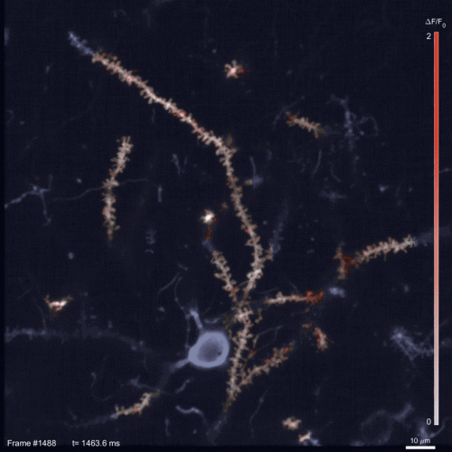
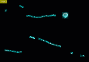
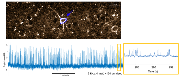
SLAP2 voltage imaging using integration scanning. Top: Blue lines outline a single integration ROI. Bottom Left, 5 minute voltage recording from the selected cell, raw intensity shown (no bleaching compensation or baseline subtraction). Inset Bottom Right, zoom in intensity trace during 5th minute of recording.
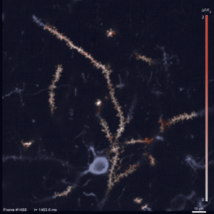
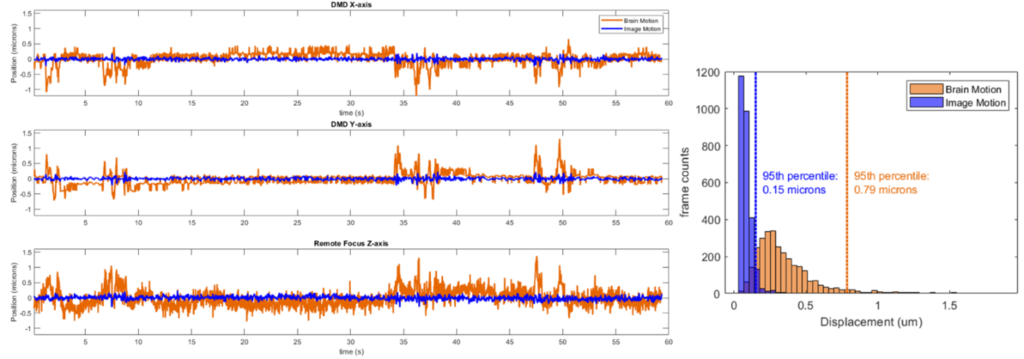
Performance of SLAP2 online motion correction. Left, The motion of the brain (orange) and the residual image motion not corrected by online motion correction (blue) over a typical 60-second period while imaging an iGluSnFR3-expressing neuron in a headfixed mouse at 220 Hz; The 3 spatial axes are shown. Right, histogram of 3D displacements for brain motion and residual image motion after online correction.
SLAP2 performs Random Access Imaging without access time costs
‘Random Access Imaging’ is the ability to record from only voxels of interest, enhancing imaging speed and excitation efficiency. Other widely0used random access scanning relies on acousto-optic deflector (AOD) crystals, which must pay an ‘access time’ cost for each new target they address. For this reason, AOD-based microscopes cannot record from large numbers of targets (i.e. neurons or synapses) at high speed. SLAP2 has zero per-target access time cost, making it far faster and more efficient for recording from large numbers of targets. This is a critical feature for synaptic activity imaging.
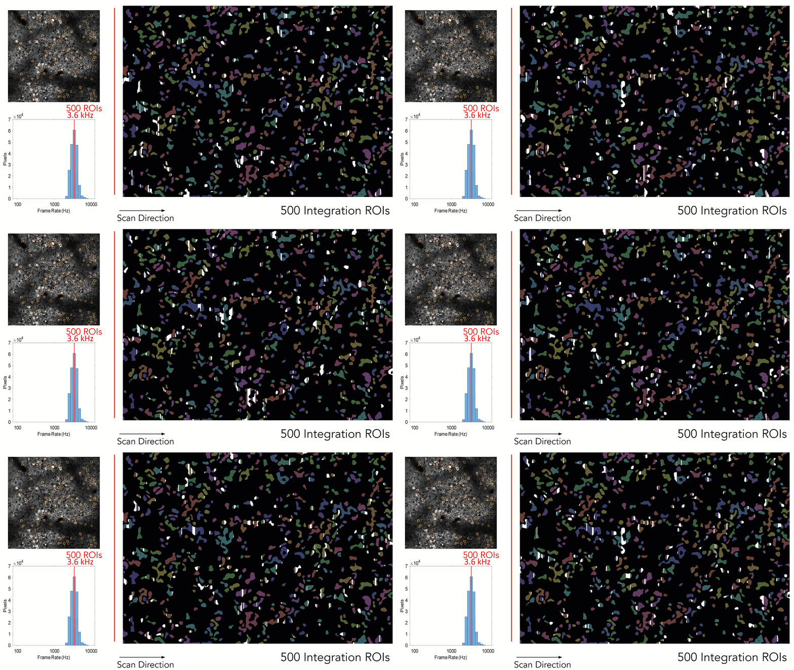
SLAP2 ‘Integration ROIs’ illuminate all pixels in a target at once, enabling multi-kiloHertz imaging
For applications such as voltage imaging, SLAP2 excites all the pixels in a neuron on each laser sweep, enabling population recordings at up to 21.6 kHz. This approach, Integration Scanning, is both faster and more power efficient than raster scanning, performing high-SNR voltage recordings using 4 mW of power per neuron in vivo. Integration scanning excites only selected pixels and illuminates them with the same intensity as point scanning, only simultaneously rather than in sequence. Because only pixels within each target are illuminated at once, there is no mixing of signals across ROIs.
SLAP2 performs fast, high-performance image-based motion correction to enable in vivo imaging
SLAP2 uses incoming imaging data to update its actuators, compensating for sample movement with a latency of <100 µs. This approach does not require the implantation of beads or definition of additional scan targets that slow down imaging. In tests with behaving mice at the Allen Institute, the 95th percentile of residual movement was 150 nanometers.
Two remote-focused imaging paths to address multiple sample depths
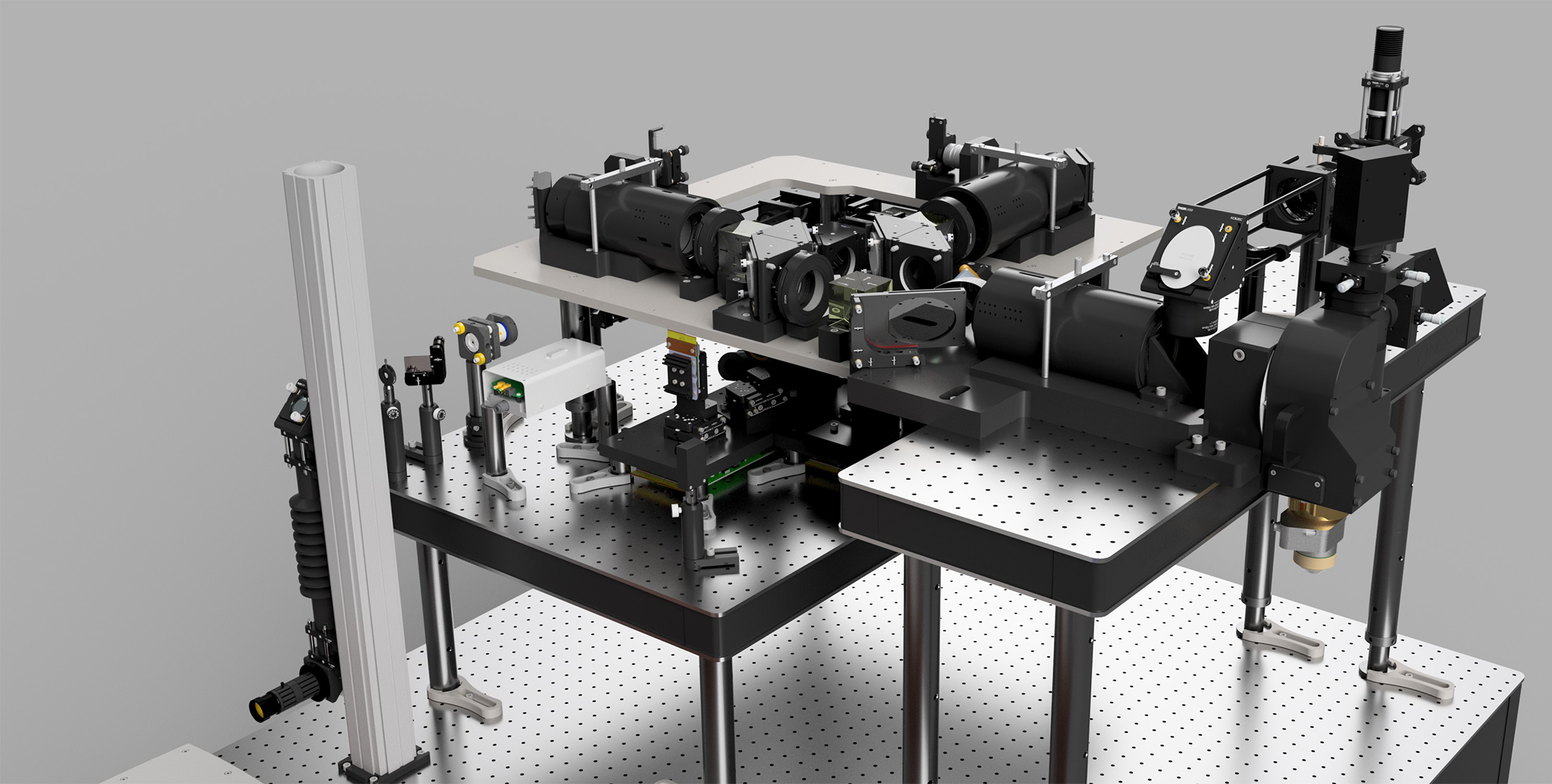
SLAP2 uses remote focusing to move each imaging path’s focus depth without moving the sample or objective. Remote focusing is extremely fast and independent for the two imaging paths.
User-friendly interface.
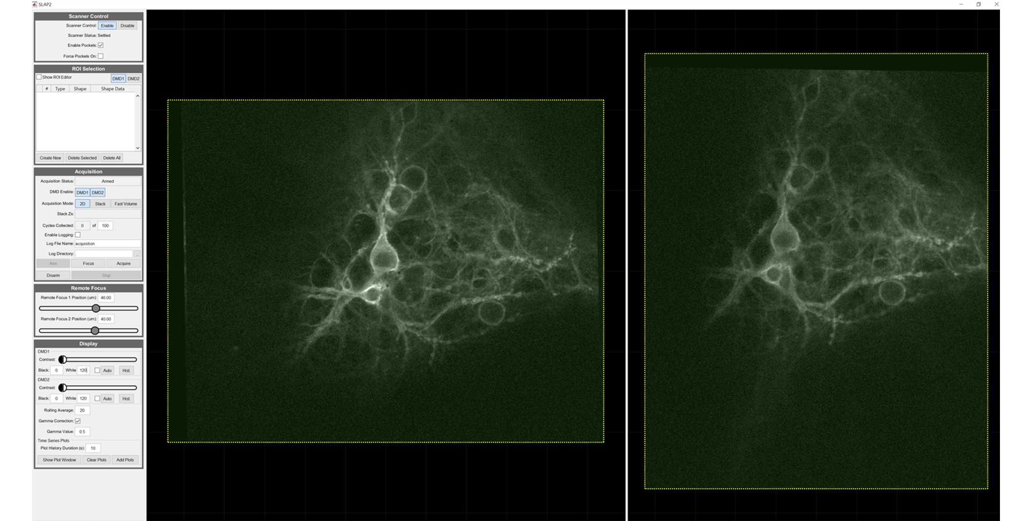
SLAP2 has a mature and full-featured GUI for defining scan and visualization parameters, interfacing with other instruments, and performing closed-loop experiments. SLAP2 shares user interface features with ScanImage.
Cited in Peer Reviewed Scientific Publications
MBF’s utility is underscored by the number of references it receives in the worlds most important scientific publications.
Boeglin, M., E. Leyva-Díaz, et al.
Expression and function of C. elegans UNCP-18, a paralogue of the SM protein UNC-18View Publication

Rentsch, P., T. Egan, et al.
The ratio of M1 to M2 microglia in the striatum determines the severity of L-Dopa-induced dyskinesiasView Publication

Wang, Z., D. Zheng, et al.
Enabling Survival of Transplanted Neural Precursor Cells in the Ischemic BrainView Publication

Villar-Conde, S., V. Astillero-Lopez, et al.
Synaptic involvement of the human amygdala in Parkinson’s diseaseView Publication

Stimpson, C. D., J. B. Smaers, et al.
Evolutionary scaling and cognitive correlates of primate frontal cortex microstructureView Publication

Russ, T., L. Enders, et al.
2,4-Dichlorophenoxyacetic Acid Induces Degeneration of mDA Neurons In VitroView Publication

Olkhova, E. A., C. Bradshaw, et al.
A novel mouse model of mitochondrial disease exhibits juvenile-onset severe neurological impairment due to parvalbumin cell mitochondrial dysfunctionView Publication

Zhang, X., C. Wang, et al.
Analysis of Error Sources in the Lissajous Scanning Trajectory Based on Two-Dimensional MEMS MirrorsView Publication

Lu, J., Behbahani, A.H., Hamburg, L. et al.
"Transforming representations of movement from body- to world-centric space."

Who Is Using MBF Products?
MBF products are used across the globe by the most prestigious laboratories.


Testimonials
"I rarely have encountered a company so committed to support and troubleshooting as MBF."

Andrew Hardaway, Ph.D. Vanderbuilt University
"MBF Bioscience is extremely responsive to the needs of scientists and is genuinely interested in helping all of us in science do the best job we can."

Sigrid C. Veasey, MD University of Pennsylvania
"I am so happy to be a customer of your company. I always get great help related with your product or not. With the experienced members, you are the best team I've ever met. All of your staff are very kind and helpful. Thank you for your great help and support all the time."

Mazhar Özkan Marmara Üniversitesi Tıp Fakültesi, Turkey
"We’ve been very happy for many years with MBF products and the course of upgrades and improvements. Your service department is outstanding. I have gotten great help from the staff with the software and hardware."

William E. Armstrong, Ph.D. University of Tennessee
"Our experience with the MBF equipment and especially the MBF people has been outstanding. I cannot speak any higher about their professionalism and attention for our needs."

Bogdan A. Stoica, MD University of Maryland
"MBF provides excellent technical support and helps you to find the best technical tools for your research challenges on morphometry."

Wilma Van De Berg, Ph.D. VU University Medical Center - Neuroscience Campus Amsterdam
Robust Professional Support
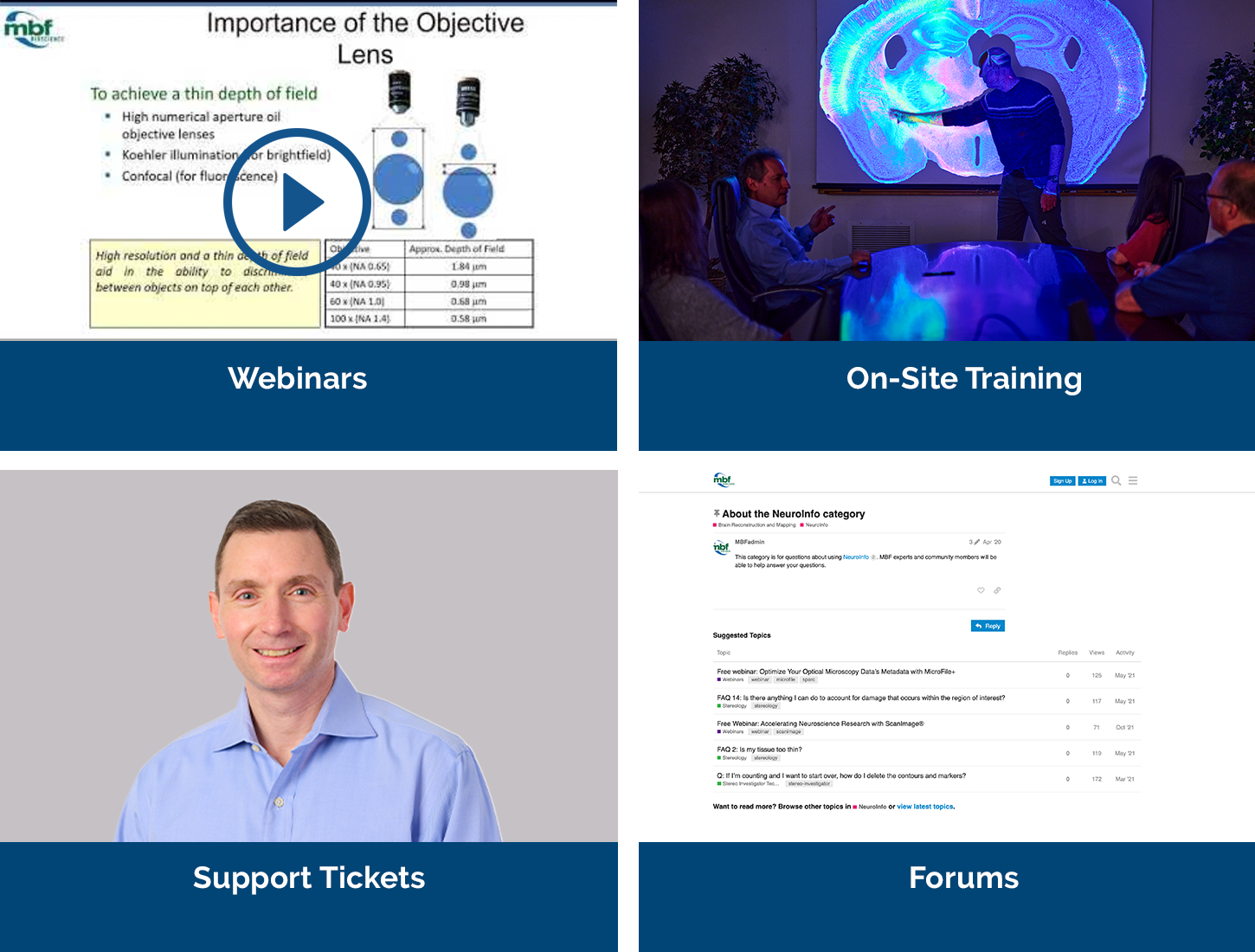
Our service sets us apart, with a team that includes Ph.D. neuroscientists, experts in microscopy, stereology, neuron reconstruction, and image processing. We’ve also developed a host of additional support services, including:
- Forums
We have over 25 active forums where open discussions take place.
>> Learn More - On-Site/Training
We’ve conducted over 750 remote software installations.
>> Learn More - Webinars
We’ve created over 55 webinars that demonstrate our products & their uses.
>> Learn More
Request more Information
If you’re interested in building your own SLAP2 microscope, MBF Bioscience has everything you need! Contact us for more details.

Related Products
vDAQTM
Vidrio’s Data Acquisition Card is a next generation all in one controller for your microscope. It controls Galvos, Pockels Cells, Fast Z devices, shutters and much more.
ClearScope® - The Light Sheet Theta Microscope
Ground-breaking light sheet microscope system for cleared specimens.


