Count objects
Optical Fractionator - step 10
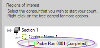
The workflow displays a list of the sections and contours that were drawn for each section.
- Click the name of a contour to select it.
- Optional: Sampling: Confirm the sampling parameters to be used.
- Click Start counting. The workflow displays new options for counting.
- The stage moves to the first counting site and, if you selected Refocus to top of section at each grid site in step 5, the Focus Top Of Section window is displayed.
- Focus above the top of the tissue until it is completely out of focus.
- Slowly focus back down onto the tissue until something on the tissue (e.g., a cell or interstitial components) comes into focus; this is the top of the section.
- Click OK.
- The Focus Bottom Of Section window is displayed.
- Focus all the way down through and past the bottom of the tissue until it is completely out of focus.
- Slowly focus back up until something just comes into focus. This is the bottom of the tissue (use the Z-meter to help you determine the direction that you are focusing.
- Click OK.
Determining what point is the top and what point is the bottom of the tissue varies from one researcher to another.
This is not an issue as long as you are the only one counting a particular experiment, and as long as you are consistent in your criteria
- Start counting in the first site.
- Select a marker from the Use Marker drop-down menu in the workflow.
- Optional: Check the Enable middle mouse button for placing markers box to mark two populations simultaneously with identical sampling parameters (use for population estimates of two similarly frequent populations).
- Focus through the tissue to find the unique point of the particle. If you focus above or below the tissue (i.e., outside of the green area displayed in the Z meter), the cursor changes to indicate that you're outside of the appropriate area.
 About the unique point
Many researchers use the cell's top, but the top of the nucleus or of the nucleolus (provided that the nucleolus is unique to the cell) is fine. The "unique point" must fall within your disector height (shown as green in the z meter). The rest of the cell may be anywhere relative to the counting frame, but only the unique point matters when deciding whether or not to mark a cell.
- If the unique point comes into focus while in the guard zone (shown as red in the z meter), do not count it!
- When the unique point comes into focus, place a marker on it in accordance with the rules of the counting frame.
- Click the "unique point" to place the marker. Counting rules for the counting frame
- Repeat steps c-d for the other particles that can be counted in the site.
- Once you've identified all the particles in the site, click the button in the workflow to move to the next counting site.
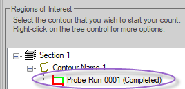
- Once you have visited all the sites, the probe run is displayed under Regions of Interest .
- Select the next section: Click the Begin Next Section button.
- The workflow redirects you to a previous step.
- The program verifies that you have the
- Some steps are disabled since the same parameters must be used for all sections in a given region.
- The program verifies that you have the
- Follow the workflow down to the Count Objects step again.
- Continue counting in each section and adding new sections until you have completed all the sections in this particular specimen.
- Click I've Finished Counting.
One file = one specimen
● DO NOT save each section as a new file.
● Use the workflow to add new sections until you've traced and counted all the sections in an animal; this allows you to save these sections as one file, allowing you to view the total cell estimate for the entire structure, not just one section of the structure.
See Additional commands to manage the probe runs for more options, How do I load images from an external image source with Optical Fractionator?