 NeuroInfo® User guide
Version 2026
Overview
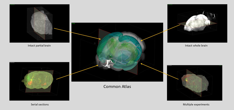
NeuroInfo enables whole-brain investigation of circuits, neuronal populations, and biochemical-marker expression using widely accepted brain atlases for anatomic reference. Visualize 2D and 3D brain microscopy images, register to a standardized reference brain, visualize and quantify brain region delineations.
What's new
See the What's new page or review the Release Notes.
Getting started
-
See an overview of the NeuroInfo software interface/workspace.
-
Review a glossary of terms used in NeuroInfo.
-
Create a 3D brain reconstruction from 2D serial-section microscopy image data using the Serial Section Assembler.
-
Detect cells and place markers at their locations using the Cell detection workflow.
-
Identify brain regions in images of your samples by comparison to a public brain atlas. Start by registering (aligning) your 3D brain images to a reference-brain atlas, using either:
-
Register Sections for 3D image reconstructions derived from serially sectioned tissue, or to register a single or multiple serial sections
-
Register Volume for brain images from tissue that was intact, i.e., not sectioned or sliced prior to imaging or to refine the registration of a reconstructed brain image volume that was registered using the Atlas Constrained Section Registration, then mapped to an intermediate space.
-
-
Once registration is complete, you can map data, such as markers for detected cells and contours that designate specific brain regions to obtain quantitative information about where the data are located.
NeuroInfo webinars on YouTube
Computer-system requirements
Guidelines on computer-system requirements are available here.
The computer-processing power needed to run NeuroInfo depends on the size, compression, and structure of the image and data files that you are evaluating. We recommend that you review the computer-system requirements before you start working with NeuroInfo or if the software crashes while you are using it.