New Therapy May Aid Heart Repair After Heart Attack
Researchers Quantify Improvement in Heart Vasculature with Vesselucida 360 and Vesselucida Explorer Cells need oxygen to survive, but during a heart
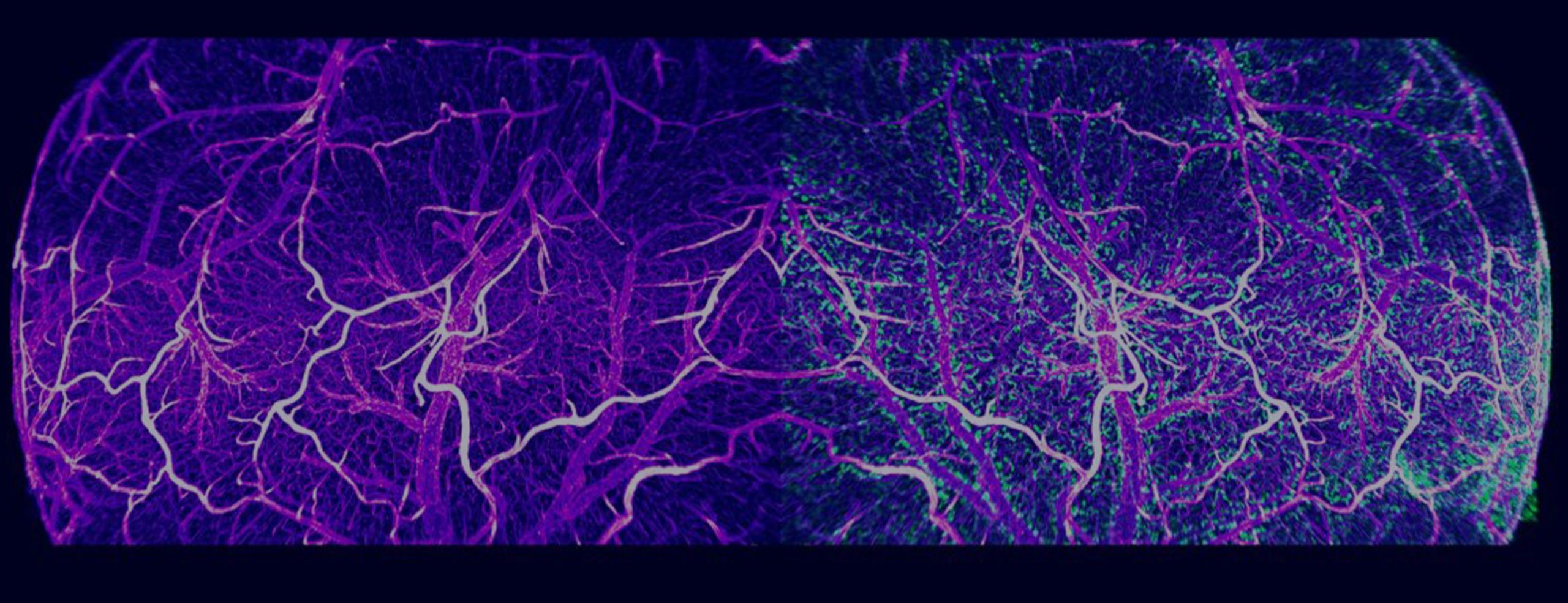
Vesselucida Explorer is the analytical software companion for Vesselucida 360 – engineered to perform extensive morphometric analyses on microvascular models and serial section reconstructions.
Dynamically visualize and comprehensively analyze vessels, puncta, and regions in an intuitive 3D environment while making quantitative measurements. Vesselucida Explorer is highly interactive, enabling researchers to explore their data models and export quantitative metrics generated directly from the models.
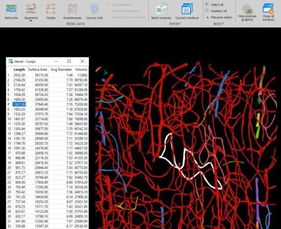
Comprehensive quantitative vascular and anatomical morphometric analysis
Understanding the vascular makeup of cerebral architecture or visceral organs is an integral part of uncovering mechanisms of disease and finding new treatments. With Vesselucida Explorer, you can quantify vascular morphology from vessel reconstruction data derived in Vesselucida 360 or Vesselucida.
Vesselucida Explorer is equipped with dozens of neuronal and regional analyses that are customizable to your specific research questions. We have removed the obstacles for rapid, high-volume, quantitative data analysis by programming all the complex morphological analyses, so you don’t have to.
| Minimum System Requirements | |
| Operating system | Windows 10, 64-bit |
| Input Specifications | |
| Supported image file formats | XML, ASC, or DAT generated in Vesselucida 360 or Vesselucida |
| Output Specifications | |
| Tracing export formats | Data model: XML*, ASC, DAT, NRX Vector: SVG Images: TIFF, JPEG, PNG Marker coordinates: TXT |
|
| * Our XML data file format, the Neuromorphological File Specification (NFS), was recently endorsed as a standard by the INCF. |
Vesselucida Helps Researchers Quantify Post-Injury Capillary Damage and Regeneration
>> Learn More
New Therapy May Aid Heart Repair After Heart Attack
>> Learn More
Scientists use Vesselucida 360 to quantify brain vasculature in mTBI model
>> Learn More
Download Vesselucida Explorer product sheet here.
Vesselucida Explorer version 2022.1.1 (Released December 2022)
New Features and Enhancements
View Full Version History Here.
Puncta analyses allow you to perform morphometric and quantitative measurements on the puncta that were detected in Vesselucida 360. The specific channels used for the detections are respected and can be used to compare the different populations of puncta to each other and to vessel structures. The analyses include area, volume, colocalization, nearest neighbor, distance to structures, and number of puncta in closed contours, closed surface and layers.
Vesselucida Explorer is used across the globe by the most prestigious laboratories.


Researchers Quantify Improvement in Heart Vasculature with Vesselucida 360 and Vesselucida Explorer Cells need oxygen to survive, but during a heart
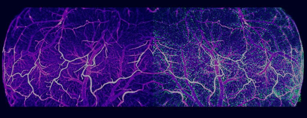
Williston, VT – September 5, 2018 – Researchers studying microvascular networks and vessels have a groundbreaking new software application to facilitate
Vesselucida Explorer’s utility is underscored by the number of references it receives in the worlds most important scientific publications. See examples below:
Buncha, V., K. A. Fopiano, et al.
Mice deficient in endothelial cell-selective adhesion molecule develop left ventricle diastolic dysfunctionView Publication

Brady, E. L., O. Prado, et al.
Engineered tissue vascularization and engraftment depends on host modelView Publication

Jacobsen, N. L., C. E. Norton, et al.
Myofibre injury induces capillary disruption and regeneration of disorganized microvascular networksView Publication

Gama Sosa, M. A., R. De Gasperi, et al.
Late chronic local inflammation, synaptic alterations, vascular remodeling and arteriovenous malformations in the brains of male rats exposed to repetitive low-level blast overpressuresView Publication

Sullivan, A.E., Tappan, S.J., Angstman, P.J. et al.
A Comprehensive, FAIR File Format for Neuroanatomical Structure Modeling. Neuroinform (2021). https://doi.org/10.1007/s12021-021-09530-xView Publication

Gama Sosa, M.A., De Gasperi, R., Pryor, D. et al.
Low-level blast exposure induces chronic vascular remodeling, perivascular astrocytic degeneration and vascular-associated neuroinflammation. acta neuropathol commun 9, 167 (2021). https://doi.org/10.1186/s40478-021-01269-5View Publication

Vesselucida Explorer software is complimentary to the purchase of Vesselucida or Vesselucida 360 software. Additional licenses of Vesselucida Explorer sold separately.
Yes. Though Vesselucida Explorer can be directly launched from Vesselucida or Vesselucida 360 applications, the software can be installed separately on other computers in your lab to increase data generation and interpretation throughput.
With a click of a button, the tabular morphometric data generated by Vesselucida Explorer can be easily exported to Excel. For researchers who prefer to program custom analyses and operations, data can be exported as separate files for loading into other software such as Matlab, Python, or R.
MBF is proud to help bioscience leaders and experts push the next wave of innovation and discovery to benefit humanity. Given this, it’s possible you’re looking for customized technology for your needs. If that is the case, click here to get in touch with us directly. We’d be happy and honored to see if we can create something together that will move all of us forward.
Vesselucida Explorer is the analytical software companion for Vesselucida and Vesselucida 360. Researchers can use Vesselucida 360 or Vesselucida to generate vessel reconstructions and anatomical models from acquired image data or directly from slides at the microscope, respectively. Then you can leave the image data and microscope behind, and employ Vesselucida Explorer to quantify the Vesselucida 360 or Vesselucida -derived models.
All MBF Bioscience software comes with comprehensive, context-sensitive help guides accessible online or offline. All of the analysis available in Vesselucida Explorer have been fully documented in the product user-guide.
"I rarely have encountered a company so committed to support and troubleshooting as MBF."

Andrew Hardaway, Ph.D. Vanderbuilt University
"Vesselucida enables us to visualize and quantify microvascular networks in a way that has not been done before. The ability to render 3D maps is integral to analysis of network structure, remodeling and blood flow distribution."

Dr. Nicole Jacobsen University of Missouri
"MBF Bioscience is extremely responsive to the needs of scientists and is genuinely interested in helping all of us in science do the best job we can."

Sigrid C. Veasey, MD University of Pennsylvania
"After examining different vessel quantification tools for use with neovessel formation in the heart, we chose to use Vesselucida 360 because it offered us flexibility in viewing and adjusting vessel geometries in 3D to accurately represent our microCT dataset. Also, the quantitative parameter extrapolation is excellent."

Dr. Kareen Coulombe Brown University
"I am so happy to be a customer of your company. I always get great help related with your product or not. With the experienced members, you are the best team I've ever met. All of your staff are very kind and helpful. Thank you for your great help and support all the time."

Mazhar Özkan Marmara Üniversitesi Tıp Fakültesi, Turkey
"We’ve been very happy for many years with MBF products and the course of upgrades and improvements. Your service department is outstanding. I have gotten great help from the staff with the software and hardware."

William E. Armstrong, Ph.D. University of Tennessee
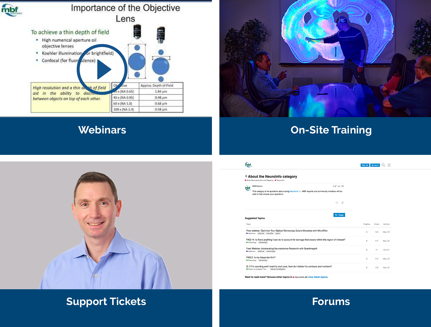
Our service sets us apart, with a team that includes Ph.D. neuroscientists, experts in microscopy, stereology, neuron reconstruction, and image processing. We’ve also developed a host of additional support services, including:
We offer both a free demonstration and a free trial copy of Vesselucida Explorer. During your demonstration you’ll have the opportunity to talk to us about your hardware, software, or experimental design questions with our team of Ph.D. neuroscientists and experts in microscopy, neuron tracing, and image processing.
