Vesalius®
Versatile spinning disk confocal microscope system ideal for fast, large-scale, multichannel 2D and 3D whole slide imagingProduct Overview
Vesalius is a versatile, cost-effective, confocal microscope system that is ideal for fast, large-scale, multi-channel 2D and 3D whole slide imaging of slide mounted specimens. Capture stunning images of thick or thin tissue samples, with customizable imaging options at speeds that are 10x faster than conventional laser scanning confocal microscopes and more than 100x faster than grid-based structured illumination. Benefit from high signal-to-noise ratio, minimal photobleaching, and eliminate out-of-focus planes, all while maximizing your productivity and budget with the fastest and most versatile confocal imaging system available.
Key Benefits
- Create high-quality large-scale 3D images: from single stacks - up to whole slides
- Use a wide range of objective lenses – from 4x up to 100x
- More than 10x faster than a traditional confocal microscope
- Custom designed and configured systems for the most cost-effective confocal solution
- Powered by our easy-to-use image acquisition software to handle big image data and confocal stereology
The Vesalius system (developed in concert with CrestOptics) is the perfect tool for fast, multi-channel confocal image acquisition of large tissue specimens, including mouse, rat and non-human primate. Vesalius is easy to use and substantially reduces the time needed for image acquisitions compared to conventional confocal imaging systems, with no compromise in image quality.
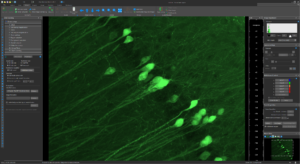
Combine MBF Bioscience award-winning image analysis software, using the latest AI technology, with your Vesalius system to perform complex quantitative image analyses such as automated cell counting, neuron reconstruction and dendritic spine detection, as well as our gold-standard stereological analysis solutions.
You can include powerful analysis tools with your customized Vesalius system:
-
- Automated cell detection and rapid stereological analysis in 2D and 3D images (Stereo Investigator® AI)
- Trusted stereological analysis (Stereo Investigator®)
- Assembly of full-resolution 3D images from 2D serial-section image data (BrainMaker®)
- Automated neuron tracing and quantitative analysis (Neurolucida 360®)
- Automated vessel tracing and quantitative analysis (Vesselucida 360®)
Vesalius can also be fully integrated with existing MBF Bioscience microscopy systems, including Stereo Investigator® and Neurolucida®. Acquire vast amounts of data from standard slides, expansion microscopy, or cleared tissue and analyze it with our software solutions that are developed for neuroscience research in collaboration with experts in the advanced-microscopy research community.
| Acquisition speed over 1,000 fps on full Field of View (25mm) |
| Axial resolution (FWHM): ~600nm depending on objective lens |
| Image Depth: up to ~200um |
| FOV Field of view up to 25mm |
| Linear encoded motorized stage for the greatest accuracy and flexibility |
| Wide range of illumination sources (laser or LEDs, up to 7 excitation wavelengths from 390nm-750nm) |
Spinning disk geometry (diamater/spacing)
|
| Resolution
|
Download Vesalius product sheet here.
-
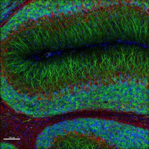
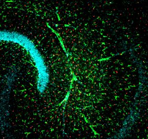
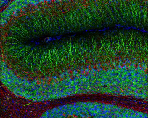
- Rat Brain cerebellum acquired with 40x/0.75 NA objective on Vesalius. Tissue was labeled with Hoechst (blue), MAP2-conjugate (green), and RPCA-NFL-ct (red). Slide prepared and provided by Encor Biotechnology Inc.
-
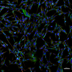
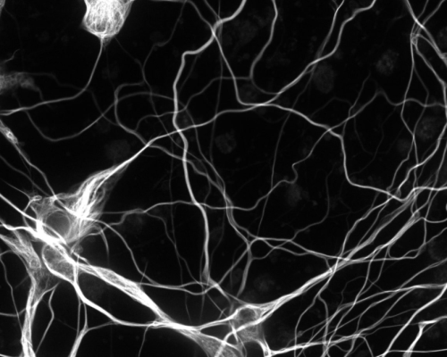
- A72 Canine Fibrosarcoma Cells acquired on Vesalius. Tissue was labeled with Hoechst (blue), Actin-phalloidin (green), and β- Tubulin (red). Slide prepared and provided by Encor Biotechnology Inc.
Vesalius: Key Features
Versatility for today’s labs and core facilities that work on a wide range of projects
- Multiple imaging modes available: Confocal, Live Cell, Widefield, Brightfield, Darkfield
- Customizable configurations include:
- Up to 7 color channels using LEDs or lasers
- Objective lenses from 4x to 100x
- XY motorized stages can hold multiple slides or XXL slides for large tissue specimens
- Fast Z-stacks, 2D whole slide imaging, and 3D whole slide imaging
Vesalius: Key Features
Value for large and small labs
- Order a new custom-configured, turn-key system specifically designed for your research
- Upgrade your existing fluorescence microscope to a state-of-the-art spinning disc confocal
- Work with state-of-the-art components, e.g., Zeiss, Olympus, Nikon or Leica microscopes (upright and inverted), ultra-sensitive high-resolution cameras, motorized stages, and multi-channel lasers
- Easy-to-use, multi-purpose system
Vesalius: Key Features
Vigorous, reproducible and reliable results
- Fast confocal imaging with scan rates of up to 1,000 fps enables you to capture more data in less time and speed up your research
- Benefit from large field of view of up to 25mm, which translates to fewer tiles for creating montages and performing whole-slide imaging
- High resolution and high sensitivity combined to create a crystal-clear confocal imaging system to help you see more from your experimental specimens.
- Reliability is built in—we use only the most robust components from trusted names in microscopy. Vesalius will be a true workhorse in your laboratory or imaging facility for many years
Interested in Upgrading your Existing Stereo Investigator or Neurolucida system?
Learn more about upgrading your existing Stereo Investigator or Neurolucida system with Vesalius for true, high-speed, high-throughput confocal stereology, and to analyze vast amounts of high-resolution data with our latest AI technology.
Learn more about how we can custom configure a new Vesalius spinning disk confocal microscope specifically for your research needs
Don’t settle for an out-of-the-box microscope that costs too much and doesn’t meet all of your needs. Maximize your budget with the most versatile system available without paying for features you don’t need.
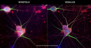
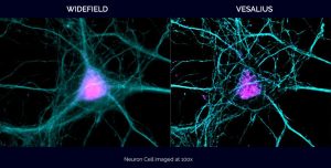
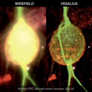
The Vesalius enables a dynamic widefield/confocal transition and true Z optical sectioning in a single click.
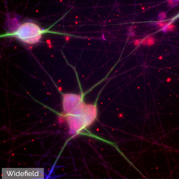
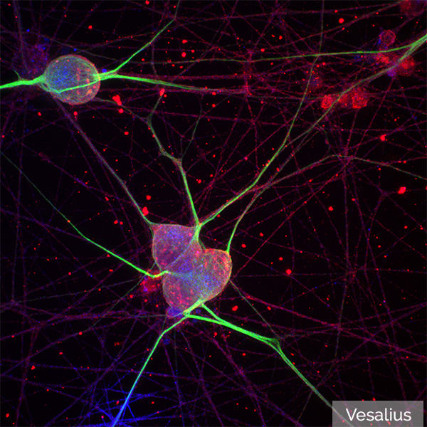
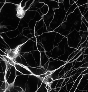
Human iPS derived motoneurons
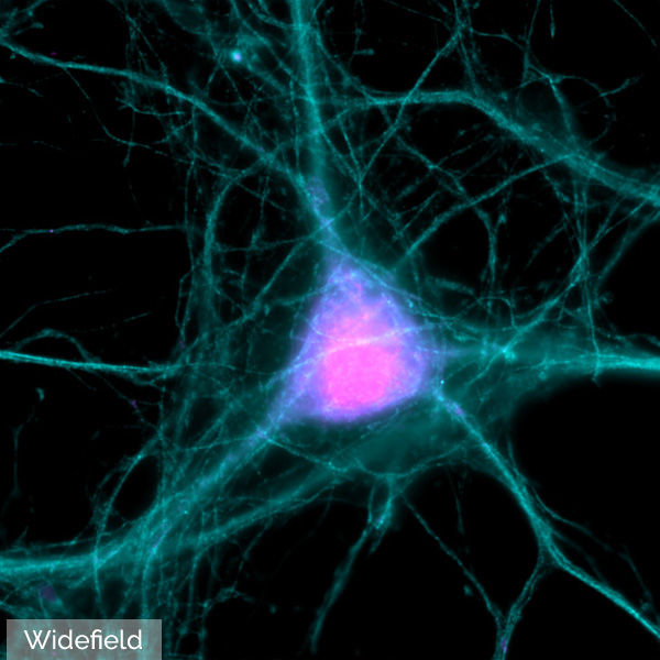
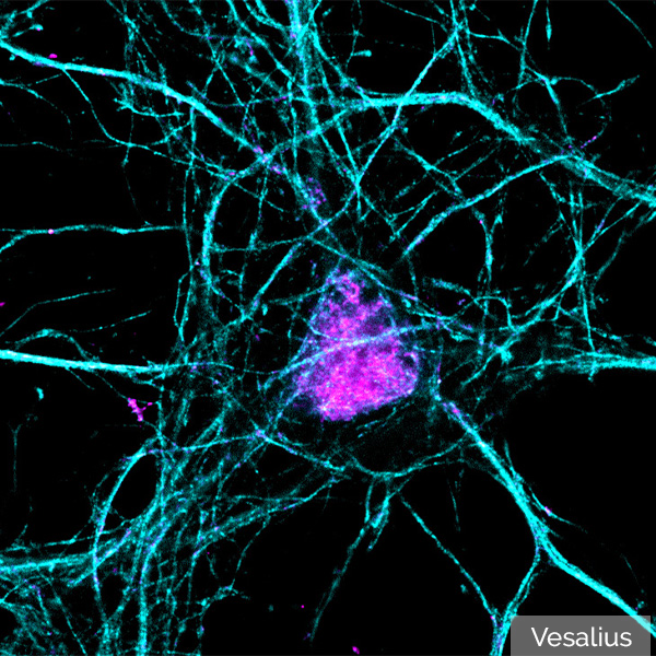
Neuron cell imaged at 100x
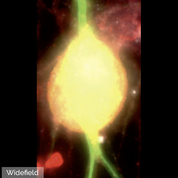
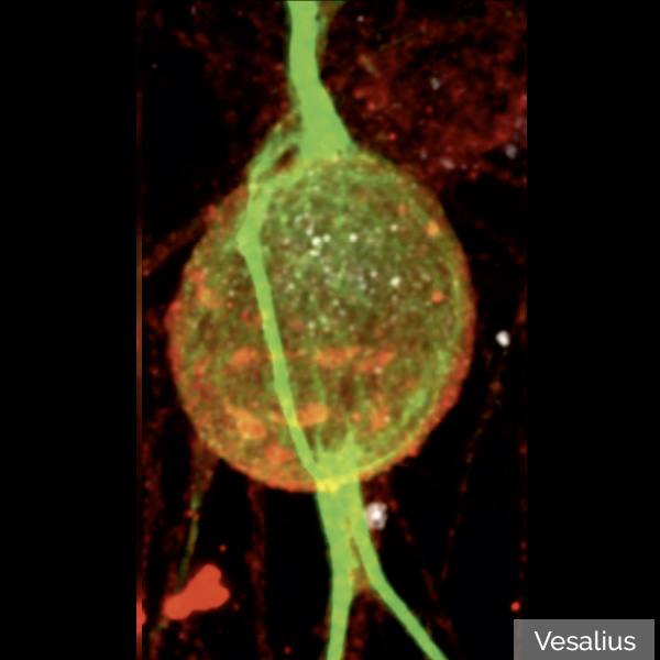
Human iPSC derived motoneurons. 60x oil
Cited in Peer Reviewed Scientific Publications
MBF’s utility is underscored by the number of references it receives in the worlds most important scientific publications.
Boeglin, M., E. Leyva-Díaz, et al.
Expression and function of C. elegans UNCP-18, a paralogue of the SM protein UNC-18View Publication

Rentsch, P., T. Egan, et al.
The ratio of M1 to M2 microglia in the striatum determines the severity of L-Dopa-induced dyskinesiasView Publication

Wang, Z., D. Zheng, et al.
Enabling Survival of Transplanted Neural Precursor Cells in the Ischemic BrainView Publication

Villar-Conde, S., V. Astillero-Lopez, et al.
Synaptic involvement of the human amygdala in Parkinson’s diseaseView Publication

Stimpson, C. D., J. B. Smaers, et al.
Evolutionary scaling and cognitive correlates of primate frontal cortex microstructureView Publication

Russ, T., L. Enders, et al.
2,4-Dichlorophenoxyacetic Acid Induces Degeneration of mDA Neurons In VitroView Publication

Who Is Using MBF Products?
MBF products are used across the globe by the most prestigious laboratories.


Testimonials
"I rarely have encountered a company so committed to support and troubleshooting as MBF."

Andrew Hardaway, Ph.D. Vanderbuilt University
"MBF Bioscience is extremely responsive to the needs of scientists and is genuinely interested in helping all of us in science do the best job we can."

Sigrid C. Veasey, MD University of Pennsylvania
"I am so happy to be a customer of your company. I always get great help related with your product or not. With the experienced members, you are the best team I've ever met. All of your staff are very kind and helpful. Thank you for your great help and support all the time."

Mazhar Özkan Marmara Üniversitesi Tıp Fakültesi, Turkey
"We’ve been very happy for many years with MBF products and the course of upgrades and improvements. Your service department is outstanding. I have gotten great help from the staff with the software and hardware."

William E. Armstrong, Ph.D. University of Tennessee
"Our experience with the MBF equipment and especially the MBF people has been outstanding. I cannot speak any higher about their professionalism and attention for our needs."

Bogdan A. Stoica, MD University of Maryland
"MBF provides excellent technical support and helps you to find the best technical tools for your research challenges on morphometry."

Wilma Van De Berg, Ph.D. VU University Medical Center - Neuroscience Campus Amsterdam
Robust Professional Support
Our service sets us apart, with a team that includes Ph.D. neuroscientists, experts in microscopy, stereology, neuron reconstruction, and image processing. We’ve also developed a host of additional support services, including:
- Forums
We have over 25 active forums where open discussions take place.
>> Learn More - On-Site/Training
We’ve conducted over 750 remote software installations.
>> Learn More - Webinars
We’ve created over 55 webinars that demonstrate our products & their uses.
>> Learn More
Request more Information
At MBF, we’ve spent decades understanding the needs of researchers and their labs — and have a suite of products and solutions that have been specifically designed for the needs of today’s most important and advanced labs. Our commitment to you is to spend time with you discussing the needs of your lab — so that we can make sure the solutions we provide for you are exactly what you’ll need. It’s part of our commitment to supporting you — before, during, and after you’ve made your decision. We look forward to talking with you!

Related Products
Neurolucida®
Neuron tracing & analysis directly at the microscope. The gold standard for neuron tracing.
Stereo Investigator®
The complete stereology solution. The gold standard for unbiased cell counting
ClearScope® - The Light Sheet Theta Microscope
Ground-breaking light sheet microscope system for cleared specimens.


