Spaceballs workflow
You can determine the length of many different types of objects (or fibers): tubules, nerve fibers, small blood vessels, surfaces, microvilli, etc.
Spaceballs provides a length estimate instead of length-density. Reporting length per region instead of length per volume is more effective because a length-density measure can't account for possible concomitant changes in volume along with length.
The probe is a sphere or hemisphere virtually embedded in the tissue, and the counted parameter is the number of profiles that transect the edge of the sphere within a defined counting region. Since the surface of a sphere is isotropic, the need for isotropic uniform random (IUR) sections is eliminated.
Spaceballs is implemented with a fractionator sampling methodology to return an estimate of total length per region. A series of sites are selected within each section by systematic random sampling. At each site, a 3D sphere of constant volume is superimposed upon the slide to represent the intersection sites to be counted. The item to be counted is a "profile," that is, the point where the fiber transects the sphere boundary. If fibers are thick, you must identify the center point of the fiber, and only count locations where the center point transects the sphere outline.
- Thick sections (significantly thicker than the diameter of the tubules/fibers to be measured)
- Structure of interest stained through the depth of the tissue section.
- Unstained tissue transparent enough to see the stained structure throughout the depth of the section.
- Thin focal planes achieved with a high magnification, high numerical aperture lens, generally oil immersion.
- A contour traced around the region of interest.
Use a high power objective lens with a high numerical aperture to focus at the top and bottom of a few sites throughout your sections to get a rough idea of the section thickness. This value can be edited later, but an approximate value is needed for setting up the 3D sampling boxes and guard zones.
You don't need to define a counting frame for the Spaceballs probe. However, the previously defined counting frame size does have an influence on the grid size if the counting frame is larger than the desired grid.
If Preview SRS Layout does not let you specify a large enough number of sampling sites, click Probes>Tools>Define Counting Frame to define a smaller counting frame.
Click Probes>Tools>Preview SRS Layout to preview the arrangement of sampling sites within the tissue section.
If there's more than one contour in the current section, an overview of the data file is presented, and the desired contour should be selected.
Once the settings have been selected, the sampling site layout is shown in the tracing window.
You can click anywhere in the tracing window to end the Preview mode.
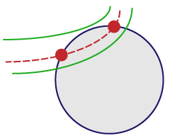
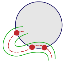
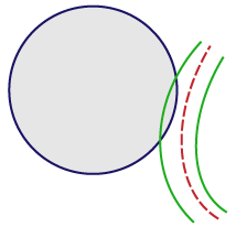
For vessels that appear on the edge of the spaceball, use the “center line” rule:
- If the center line enters and then leaves the probe, mark it twice (at the entrance point and at the exit point).

- If the center line weaves in and out, mark the centerline very time it enters or leaves the spaceball.

- If the center line doesn't enter the spaceball, don’t mark it.

- Go to Probes>Common>Spaceballs workflow.
- You are prompted to start a new subject (Start a new subject) or to continue with a subject (Load subject data from existing file).
If you select Load subject data from existing file, typically directs you to the Count Objects step. Because the workflow is streamlined, only the relevant steps are displayed and they are re-numbered accordingly (i.e., Step 10 in the complete workflow might be re-numbered Step 4 in the streamlined workflow).
- Follow the steps in the workflow.