Slide Loader setup 11: Define Virtual Tissue acquisition settings
 Define the parameters for the 3D Virtual Tissue acquisition.
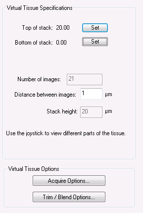
Virtual Tissue acquisition settings
Focus through the tissue to set the top and the bottom of the stack.
You need to determine a stack thickness that will minimize both the effect of tissue variation and the amount of non-usable tissue acquired.
![]() If you set up the post-load position to be focused on the top of the slide with the high power objective, the bottom of the tissue will be very close to focus position 0. If you acquire all the way through the tissue, the bottom of the stack should be near 0 as well.
If you set up the post-load position to be focused on the top of the slide with the high power objective, the bottom of the tissue will be very close to focus position 0. If you acquire all the way through the tissue, the bottom of the stack should be near 0 as well.
Virtual Tissue Options
Options available for 2D and 3D:
Keep Image Open: Displays the image in the Image Organizer when the acquisition is complete for immediate inspection.
Multichannel Acquire: Uses the existing Multichannel acquire setup; adjust the Multichannel Acquire settings prior to performing a Virtual Tissue acquisition.
Compress Tile Files: Reduces the size of the image files.
- Use the scroll bar to adjust the compression.
- Generally a compression of 10:1 or 20:1 is adequate (meaning a compression of 20:1 will take a 100 MB image file and compress it to around 5 MB).
Options available for 3D only:
Remove Temp Files: Individual image files are saved in a folder until they are compiled into the final image montage at the end of the acquisition.
- Check Remove Temp Files to delete the individual files once the image has been compiled (this will free up disk space).
Postpone Compilation: Individual image files are acquired and saved in a designated folder, but there is no compilation performed.
- Uncheck Keep Image Open and Remove Temp Files to enable the Postpone Compilation option.
- Use the Virtual Tissue Compiler when you are ready to create the montage.
Capture Virtual Tissue: Enter the distance in microns if the image needs to be acquired below the focus map.
![]() Useful if you want to create the focus map at the top of the tissue, but capture the image a set distance into the section thickness.
Useful if you want to create the focus map at the top of the tissue, but capture the image a set distance into the section thickness.
- When you designate a distance to acquire below the focus map, the first plane of the stack is acquired starting at that specified distance.
- If you enter “0”, acquisition will occur at the designated focus map locations, or at the specified top of stack.
Pixel Trim Images
Removes rows and columns of pixels from the edges of each field of view (image tile).
This option allows you to correct for spherical aberration or a video card that acquires with a black or white strip on any of the image edges.
Try a test acquisition with all of these values set to zero, and increase them if there is a problem in the final image.
- Acquire a single image
- Zoom in on the top left and bottom right corners. Any rows or columns of bad pixels are immediately evident.
- Set the trim values to remove the unwanted pixels.
For low magnifications, 0-10 pixels is a suitable range.
For higher magnifications, larger trims (25-50 pixels) may be appropriate.
- If lighting is very uneven across the field of view, large numbers of pixels may need to be removed to account for unevenly lit portions of the image
- Try to correct lighting before resorting to trimming.
- When many pixels are trimmed, the fields of view in the resulting virtual slide are made smaller, and smaller stage movements are made accordingly.
- High values for trimming will slightly increase your acquisition time, since more acquisition sites are required.
Seam Blending
Blends the edges from each field of view smoothly.
- Use the 3x3 test first to see if your image looks acceptable without blending (try to avoid blending as it involves a slight loss of image detail).
- Select the number of pixels to blend at each seam. Generally, 5-10 pixels is sufficient.
Background Color
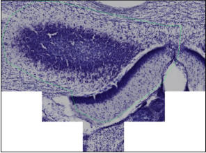
Use if the image scan selected is Contour (only inside).
- The color (White or Black) will appear around the edges of the image (white in this example:
 ).
). - Generally, white is used for brightfield imaging while black is used for fluorescent imaging.
The use of autofocus is optional.
Minimum energy
Energy is a measure of contrast in the image. In general, as the image comes into focus, the energy value increases, and the image with the highest energy corresponds to the best focus.
- If there is very little in the image (e.g., a hole in the tissue), the energy values tend to be low because there is not much contrast. For this reason, you may want to avoid that area as a focus location.
- If the autofocus is done in a region for which the best (highest energy) focused image has an energy value below the Minimum energy value that you defined, the software discards the autofocus result but maintains the Z value of the region until it can identify another region that has more to focus on.
- Use the Get button to view the energy value of the region displayed in the window. We suggest that you drive the stage to a location that doesn't contain much tissue to get an estimate of a possible minimum energy value.
 Remove slide from stage
![]() See Slide Loader Virtual Tissue Setup Workflow
See Slide Loader Virtual Tissue Setup Workflow
Stereo Investigator 11 | MBF Bioscience Support Center | Downloads