Fractionator
|
|
Number |
This probe is most often used to estimate population counts of 2D specimens such as plated cells for tissue culture analysis. Typically employed to sample populations that are too large to count exhaustively.
|
|
|
|
Mono-layer |
Performing a Fractionator probe run
- Place a reference point; the point can be placed arbitrarily or at a particular location on the slide (see Reference point overview).
- Select a low magnification lens that allows for the efficient and accurate definition of the region of interest (e.g., 4x).
- Trace a region of interest using the contour tool.
- Switch to an objective and lens that allow you to accurately identify the cells to be counted (e.g., 40x, or 20x for a sparse population).
- Click Probes>Define Counting Frame to set the size of the Counting Frame.
- Size the counting frame so that it contains a number of cells that you can comfortably mark without errors (i.e., no double count or missing cell).
- Click Probes>Preview SRS Layout to define the Systematic Random Sampling (SRS) Grid.
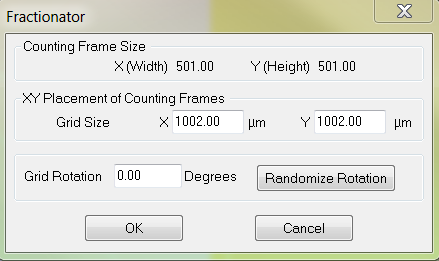
- Click Probes>Fractionator to start Fractionator.
- Stereo Investigator prompts you to open the Serial Section Manager. Click No.
- The Fractionator dialog box appears, displaying the previously determined counting frame size and grid size
 . Click OK move the stage to the first sampling site (the first site is determined randomly).
- Select a marker from the Markers toolbar and mark all the cells that fall within the counting frame following the counting rules. Counting rules for the counting frame
- When finished marking, press F2 to move to the next site.
- When finished with all the sites, the probe ends.
- To view the results, click Probes>Display Probe Run List. The Previous Stereological Runs dialog box appears.
- Select the Fraction probe for each contour (each contour represents 1 coverslip).
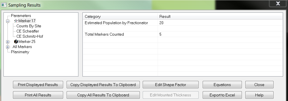
- Click View Results. The Sampling Results window appears
 .
.- Under Parameters, highlight a marker name to see the marker results on the right (estimated cell population for the coverslip).
- Highlight CE Scheaffer or CE Schmitz-Hof to see the coefficient of errors in the right panel; the values are more applicable to cell estimations in tissue sections than to coverslips. The CE can give you an indication of the homogeneity of the cellular distribution on the coverslip.See Coefficients of Error
- Highlight Planimetry to see the area of the coverslip which can be used to calculate cell density (value in the right panel).
If you don't see Planimetry, use the Options>Stereology Preferences>Results tab and check Display Planimetric Results.
![]() If the cells are double- or tripled-labeled, use combination markers to count the different cell types. See Working with Combination Markers.
If the cells are double- or tripled-labeled, use combination markers to count the different cell types. See Working with Combination Markers.
Optional: if you have multiple regions, move to the next region and repeat steps 7-10 with appropriate sampling parameters for the region.
![]() To end a Fractionator probe before it is completed, uncheck Probes>Fractionator at any time during the scan.
To end a Fractionator probe before it is completed, uncheck Probes>Fractionator at any time during the scan.
Stereo Investigator 11 | MBF Bioscience Support Center | Downloads