Deconvolution overview

Using the Huygens algorithm, the Deconvolution module reverses the optical distortion that takes place in an optical microscope to create clearer images. It can sharpen images that are affected by fast motion or jiggles during acquisition, or images that have some type of noise introduced into the signal.
How can I improve the deconvolution process?
For a typical oil lens with a numerical aperture of 1.4, use a step size no larger than 0.5 microns (0.2 is optimal).
For other lenses, consult us so that we can calculate a recommended step size based on the lens specifications.
If possible, set the pinhole on the confocal to approximately 1 airy diffraction width. Your goal is to obtain data with the best resolution possible to optimize the deconvolution process.
Remember that as you open the pinhole (usually to get more signal), axially resolution degrades. On the other hand, fluorescent signal from the specimen often requires collecting as much light as possible; it’s very common trade off.
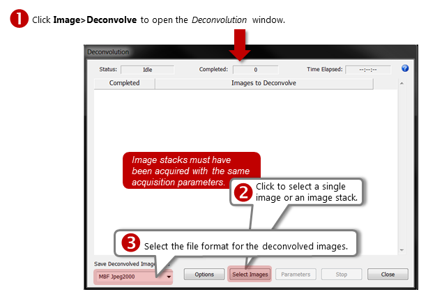
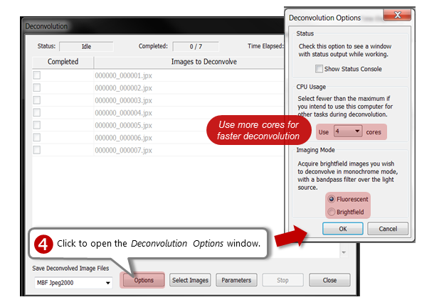
How do I use deconvolution?
All images should be closed before you start. Parameters are described below the slideshow.
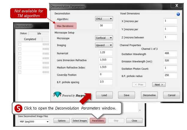

Tell me more about the Deconvolution Parameters
For confocal imagery:
- QMLE: 10-20 iterations are usually enough.
- CMLE: 30-60 iterations are appropriate. This algorithm is the most statistically rigorous, but slower.
For a representative image stack, we recommend that you to try both algorithms.
Numerical—Refers to the numerical aperture, or the amount of light coming from the focus that the objective collects.
Lens Immersion Refractive Index—Also known as the Lens Refractive Index. Refractive index of the medium (i.e., air, oil, water) the objective is immersed in.
Medium Refractive Index—Usually the same as the Lens Immersion Refractive Index. Refractive index of the medium in which the specimen is embedded.
Coverslip Position—Distance between the coverslip and the first focal plane (in Z).
B.P. pinhole spacing—Distance between the pinholes on a Nipkow disk B.P., or backprojected. Refers to the size of the pinhole as it appears on the specimen plane.
Excitation Wavelength—Wavelength of the radiation used to fluoresce the object.
Emission Wavelength—Radiation emitted back from the object.
Excitation Photon Count—Number of photons of a given excitation wavelength absorbed by a fluorophore in the object.
B.P. pinhole radius—Radius of the pinhole as seen from the focal plane. For assistance in calculating this value, see the backprojected confocal pinhole calculator page on the SVI website.
![]() Deconvolution doesn't correct highly auto-fluorescent background signal (tissue, culture media, etc.) because the deconvolution algorithms interpret the background as a real signal (i.e., as similar to all the other light intensities collected). As a result, the background is deconvolved along with the relevant experimental signal.
Deconvolution doesn't correct highly auto-fluorescent background signal (tissue, culture media, etc.) because the deconvolution algorithms interpret the background as a real signal (i.e., as similar to all the other light intensities collected). As a result, the background is deconvolved along with the relevant experimental signal.