Cleared Tissue and Whole Organ Imaging & Analysis

MBF Bioscience > Cleared Tissue and Whole Organ Imaging & Analysis

Exploring Biological Structures with Cleared Tissue Imaging


Imagine being able to see through an entire mouse brain, witnessing the intricate dance of neurons and blood vessels in perfect clarity, or peering deep inside a human tumor to understand its unique architecture. What sounds like science fiction is now a powerful reality thanks to two revolutionary techniques: tissue clearing and light sheet microscopy.


Breaking Through the Barrier of Opacity


Our bodies, and those of all animals, evolved to be opaque for good reason. Tissues are naturally packed with lipids – fatty molecules that help cells maintain their structure and function. While essential for life, these lipids scatter light, making it impossible to see deep inside organs with traditional microscopy. For over a century, scientists were limited to studying thin tissue slices, trying to reconstruct three-dimensional understanding from two-dimensional snapshots.


But what if we could make tissue transparent while keeping its structural integrity intact?

The Clearing Revolution


This is exactly what tissue clearing achieves. Think of a piece of tissue like a dense forest – light can’t penetrate very far because it’s blocked by countless obstacles. Tissue clearing is like carefully removing the foliage while keeping every tree trunk and branch exactly where it was, then filling the space with something that lets light pass through unimpeded.


Different clearing methods accomplish this in unique ways:

      • CLARITY embeds the tissue in a supportive hydrogel scaffold before removing the lipids
      • iDISCO uses organic solvents to remove lipids and homogenize the tissue’s refractive index
      • CUBIC employs gentle detergents and simple chemicals to achieve transparency
      • SHANEL specifically tackles the challenges of human tissue


Each method has its strengths, much like different tools in a toolbox. A neuroscientist studying mouse brain circuitry might choose CLARITY for its excellent structural preservation, while a cancer researcher examining human tumors might opt for SHANEL.

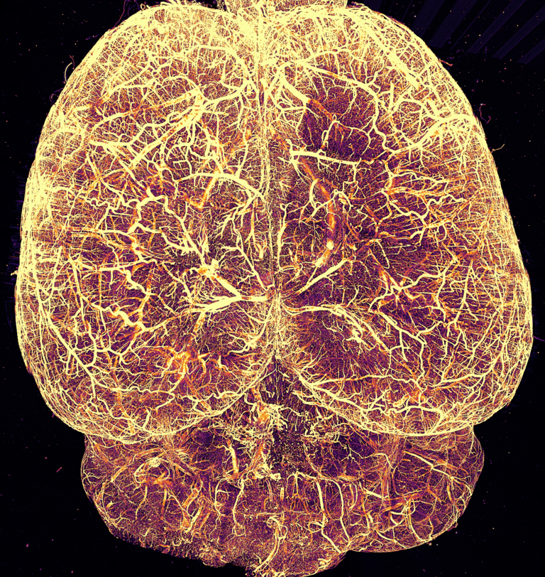
Image: High-resolution imaging of the mouse brain vasculature in an iDISCO cleared sample. Courtesy Dr. Raju Tomer, Columbia University.

Shining New Light on Biology


Making tissue transparent was only half the battle. Traditional microscopes weren’t designed to image large cleared samples effectively. Enter light sheet microscopy – a technique that transformed how we illuminate and observe cleared tissues.


Instead of shining light through the entire sample as in traditional microscopy, light sheet microscopy illuminates just one thin plane at a time with a sheet of laser light. Imagine taking a flashlight and turning its beam into a thin, flat sheet of light – that’s essentially what light sheet microscopy does. This approach brings several remarkable advantages:


      • Gentle imaging that minimizes photobleaching
      • Rapid acquisition of large volumes
      • Excellent resolution in three dimensions
      • Ability to image large samples intact


Fields Of Study

  • Connectomics
  • Neuropathology
  • Neurodegeneration
  • Proteomics
  • Expansion Microscopy
  • Vasculature
  • Cancer research
  • Neuroscience
  • Cell Biology

From Theory to Practice: Real-World Applications


The combination of tissue clearing and light sheet microscopy has opened new frontiers across biological research:


In Neuroscience

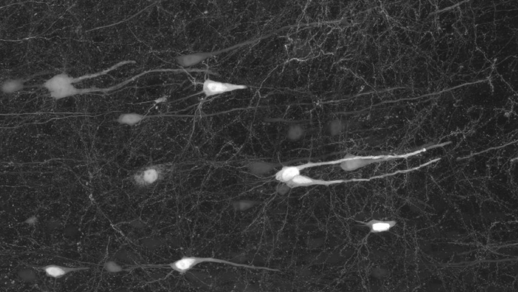
Researchers can now trace neural circuits through entire brains, mapping connections that were previously impossible to visualize. This has led to groundbreaking discoveries about how different brain regions communicate and how neural networks process information.


In Cancer Research

Scientists can examine entire tumors and their surrounding environment, understanding how cancer cells interact with blood vessels and immune cells in three dimensions. This provides crucial insights for developing more effective treatments.


In Developmental Biology

Watching how organs form and tissues develop has taken on new clarity. Researchers can observe the intricate processes of embryonic development with unprecedented detail.


The Technical Dance


Successfully imaging cleared tissue requires careful choreography of sample preparation, clearing, labeling, and imaging. Each step builds on the previous one:


      1. Preparation: The journey begins with careful fixation to preserve the tissue’s structure.
      2. Clearing: The chosen clearing method transforms the opaque tissue into a transparent window.
      3. Labeling: Fluorescent markers highlight specific structures of interest.
      4. Imaging: Light sheet microscopy captures the three-dimensional architecture.


Looking to the Future


As these techniques continue to evolve, we’re seeing exciting new developments:


      • Artificial intelligence helping to analyze the massive datasets
      • New clearing methods optimized for different tissue types
      • Advanced light sheet systems with better resolution and speed
      • Novel labeling strategies for highlighting multiple targets


The Power of Seeing More


What makes tissue clearing and light sheet microscopy so powerful is not just the ability to see deeper into tissues, but the new questions this ability lets us ask. We can now study biological processes in their natural three-dimensional context, understanding spatial relationships that were previously hidden from view.


Whether you’re a neuroscientist mapping brain circuits, a cancer researcher studying tumor development, or a cell biologist exploring tissue architecture, these techniques offer unprecedented windows into the microscopic world. They remind us that sometimes the most significant scientific advances come not from seeing something entirely new, but from seeing familiar things in entirely new ways.


Complete Workflow Solutions for Cleared Tissue Analysis


After capturing your cleared tissue images, the real work of discovery begins. MBF Bioscience provides a comprehensive suite of software tools designed specifically for analyzing complex 3D datasets from cleared tissue. Each tool serves a unique purpose in your research workflow:


NeuroInfo®: Atlas-Based Analysis Made Simple

Transform your large-scale imaging data into quantifiable insights with NeuroInfo, our advanced solution for brain-wide analysis.


Key Capabilities

      • Automated registration to standard brain atlases
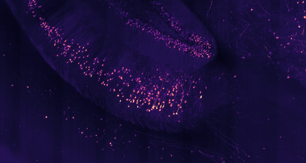
      • AI-powered cell detection across entire organs
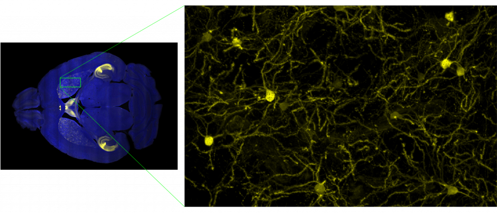
      • Region-specific cell counting and distribution analysis
      • Multi-channel support for complex labeling schemes


Perfect For

      • Whole-brain cell mapping studies
      • Activation pattern analysis
      • Brain-wide quantitative studies
      • Large-scale neuroanatomy research


How It Works

NeuroInfo seamlessly aligns your cleared tissue data with standard brain atlases, automatically identifying key brain regions and quantifying cells or markers within each area. This transforms months of manual analysis into automated workflows that complete in hours.

Neurolucida® 360: Deep Insight into Neural Architecture

The gold standard in neuron reconstruction and analysis, Neurolucida 360 brings powerful capabilities to cleared tissue research.


Key Capabilities

        • Automated neuron tracing in 3D
        • Complex morphological analysis
        • Advanced vessel tracking and analysis
        • Comprehensive morphometric analysis


Perfect For

        • Detailed neuron morphology studies
        • Vascular network analysis
        • Circuit reconstruction projects


How It Works

Neurolucida 360’s AI-powered tools automatically detect and trace neurons, spines, and vessels in your cleared tissue data. Sophisticated algorithms analyze morphology, providing detailed metrics about branch patterns, spine distributions, and network connectivity.

Stereo Investigator®: Quantitative 3D Analysis

Turn qualitative observations into quantitative data with the industry standard in stereological analysis.


Key Capabilities

        • Unbiased stereological analysis in 3D
        • Region-specific cell counting


Perfect For

        • Population studies in cleared tissue
        • Regional density analysis
        • Volumetric measurements
        • Quantitative comparison studies


How It Works

Stereo Investigator applies proven stereological principles to 3D datasets, ensuring unbiased quantification of cellular populations and structures throughout your cleared tissue samples.

NeuroDeblur®: Enhance Your Image Clarity With Deconvolution

Optimize your cleared tissue images with our advanced deconvolution software designed specifically for light sheet microscopy data.


Key Capabilities

        • GPU-accelerated processing
        • Light sheet-specific algorithms
        • Multi-view fusion
        • Batch processing capabilities


Perfect For

        • Improving image resolution
        • Reducing background noise
        • Enhancing signal-to-noise ratio
        • Processing large datasets efficiently


How It Works

NeuroDeblur uses sophisticated algorithms optimized for light sheet microscopy to remove blur and enhance detail in your images, revealing fine structures that might otherwise be hidden.


Integrated Workflow Solutions


The true power of MBF Bioscience’s tools lies in their seamless integration:


1. Acquisition to Analysis

    • Capture images with SLICE
    • Process with BrightSlice
    • Analyze with our suite of tools

2. Flexible Workflows


3. Data Sharing

    • Common data formats
    • Shared analysis parameters
    • Integrated results export

Real-World Applications


Whole Brain Analysis


    1. Acquire brain-wide images with SLICE
    2. Process and align with NeuroInfo
    3. Analyze specific neurons with Neurolucida 360
    4. Quantify populations with Stereo Investigator

Vascular Studies


  1. Image cleared vessels with SLICE
  2. Enhance vessel contrast with NeuroDeblur
  3. Trace and analyze with Neurolucida 360
  4. Quantify with Stereo Investigator

Cell Distribution Studies


  1. Capture multi-color data with SLICE
  2. Register to atlas with NeuroInfo
  3. Count and analyze with Stereo Investigator
  4. Export region-specific statistics

Support for Your Success


Expert Training


  • Personal consultation
  • Workflow optimization
  • Custom protocol development
  • Regular workshops and webinars

Technical Support


  • Direct access to experts
  • Rapid response times
  • Solution-focused assistance
  • Regular software updates

Research Community


  • User forums
  • Method sharing
  • Protocol exchange
  • Collaboration opportunities

Start Your Journey

Every research question is unique. Our team of scientists is ready to help you determine the optimal combination of tools for your specific needs. Want to explore how tissue clearing and light sheet microscopy could advance your research? Contact us to discuss your specific applications and needs.