
Neurolucida Software Update: Version 2024.1.3
We are pleased to announce the release of Neurolucida®version 2024.1.3, which introduces key updates to offline software licensing. These updates

The Neurolucida system is specifically designed for accurately tracing neuronal structures directly from histological specimens and generating quantitative morphometric data. Neuroscientists around the world trust Neurolucida as the gold standard for producing accurate anatomical models that can be quantitatively analyzed in hundreds of different ways to address perplexing biological questions. With over 6000 citations, research conducted with the help of the Neurolucida system has led to hundreds of advances in the understanding of neurodegenerative diseases, neuropathy, memory, and behavior.
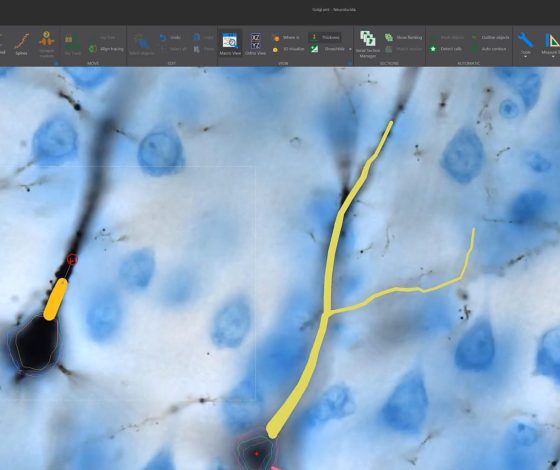
The Neurolucida system is widely recognized by neuroscientists as the gold standard for neuron tracing and reconstruction because it is based on the ground truth—you trace directly from the specimen on the microscope or from previously captured images. The tracing tools are specialized for neuronal structures, making it clear which tool to use and why; in addition, you can also edit traced structures as needed. Neurolucida offers a wide array of other powerful capabilities, including anatomical region mapping, 3D serial section reconstruction, automatic cell detection, and slide scanning.
Neurolucida fully integrates with your microscope system hardware to control motorized stages, focus, cameras, objective changers, and optical filters. Whether you need a new microscope system or want to use Neurolucida with your existing microscope hardware, our technical sales specialists will work with you to understand your current and future research interests, and choose microscope components that will help you achieve your research goals and fit your budget.
Neurolucida has been developed with support from the National Institute of Mental Health (NIMH)
| Minimum System Requirements | |
| Operating System | Windows 10, 64-bit |
| Processor | 4-core |
| CPU RAM | 16 GB |
| GPU RAM | >6 GB |
| Recommended System Requirements | |
| Operating System | Windows 10, 64-bit |
| Processor | 8-core |
| CPU RAM | 64 GB |
| GPU RAM | >8 GB |
| Storage | Solid state drive(s) |
| File Input Options | |
| Supported image file formats | CZI, HDF5, H5, IMS, JP2, JPG, JPEG, JPF, MJC, JPX, LIF, LSM, ND2, OIB, OIF, TIF, TIFF, SVS |
| Output Specifications | |
| Data output formats | XML*, ASC, DAT |
|
| * Our XML data file format, the Neuromorphological File Specification (NFS), is endorsed as a standard by the INCF. |
| Image file output formats | JP2, JPX, TIFF, SVG |
| Movie export format | MP4 |
| Hardware Compatibility | ||
|
Computer controlled microscopes | Leica supported microscopes:
| Zeiss supported microscopes:
Nikon supported microscopes
Olympus BX and IX Series:
|
| Manual microscopes | Most models after 2002 of Zeiss, Olympus, Nikon, Leica | |
|
Structured illumination |
| |
| Filter wheels and shutters | Zeiss, Olympus, Leica, Nikon, LEP, Prior, Sutter | |
|
Color video cameras |
| |
|
Monochrome video cameras |
| |
| Light sources |
| |
Supported Image File Formats: PDF
Contact us for an up-to-date list or question about specific models
Neurolucida helps to reveal new findings in a follow-up to a 40 year old study
>> Learn More
Researchers Observe Altered Neuropathology in CTE Brains
>> Learn More
Scientists Discover New “Rosehip” Neuron in Human Brain
>> Learn More
NeuroMorpho.Org Releases Nearly 10,000 New Neuron Reconstructions and Neurolucida leads the way
>> Learn More
Exercise changes astrocytes and eases symptoms of neurodegenerative disorders
>> Learn More
Uncovering the role of microglia in fetal alcohol spectrum disorders
>> Learn More
Scientists Observe Differences Between Brains of Stressed and Unstressed Rats After Fear Conditioning
>> Learn More
Hippocampal Neurons Change After Melatonin Injection
>> Learn More
Dendritic Spine Loss Reported in Schizophrenia and Bipolar Disorder
>> Learn More
Two 2014 Nobel Laureates Used Neurolucida to Create 3D Reconstructions of Neurons That Help Rats and Humans Navigate the World
>> Learn More
Anorexia Accelerates the Development of the Rat Hippocampus
>> Learn More
Scientists Use Neurolucida to Create 3D Reconstructions of Placental Villous Trees
>> Learn More
New Neurons Erase Memories
>> Learn More
Brain Cleans Itself During Sleep; Scientists Image Cerebral Fluid Flow With Neurolucida
>> Learn More
3D Reconstructions of Neurons Reveal More Branching in Sedentary Rats
>> Learn More
Neurons in the Basal Forebrain are Exquisitely Organized
>> Learn More
Scientists Discover Anorexia-Driven Changes to Dendrites With Neurolucida
>> Learn More
Neurolucida Helps Scientists Discover that Gorillas are Relevant in the Study of Alzheimer’s Disease
>> Learn More
Ohio State Neuroscientists Use Neurolucida to Analyze Brain Cells in Sexually Active Hamsters
>> Learn More
Japanese Researchers Develop New Optical Clearing Agent; Neurolucida Used For 3D Imaging in Study
>> Learn More
Drug for Treating Asthma Improves Cognitive Function in Down Syndrome Mouse Model
>> Learn More
Neurolucida Helps Ohio State Scientists Study Melatonin’s Effects on Brain Plasticity in Mice
>> Learn More
Researchers at the University of Michigan Analyze Spine Density in Addiction-Prone Rats with Neurolucida
>> Learn More
Scientists Use Neurolucida in Study of Calcium Signaling During Spontaneous Brain Activity
>> Learn More
Scientists in Portugal Use Neurolucida Explorer to Analyze Neuroplasticity in Depression
>> Learn More
Cincinnati Scientists Use Neurolucida in Epilepsy Study
>> Learn More
Michigan Scientists Analyze Brain Cells Born During Puberty With Neurolucida
>> Learn More
Stanford Scientists Render Mouse Brain Transparent, Offering New Possibilities For 3D Analysis
>> Learn More
Scientists Use Neurolucida in Study Describing New Cell Population in Juvenile CA3 Hippocampus
>> Learn More
Scientists in Japan Identify Two Brain Circuits Involved in Image Recognition; Neurolucida Plays Part
>> Learn More
UVA Scientists Use Neurolucida in Study Identifying Two New Circuits in Rat Neocortex
>> Learn More
Neurolucida Helps Scientists Map Rett Syndrome’s Brain Dysfunction in Mouse Model
>> Learn More
Scientists Map Diaphragm’s Motor Nerve and Arteriolar Networks With Neurolucida
>> Learn More
Neuroscientists in Germany Outline Protocol for Best Accuracy in Neuron Reconstruction, including Use of Neurolucida
>> Learn More
Gene Therapy May Be Answer to Effective Parkinson’s Treatment; Neurolucida Plays Role in Study
>> Learn More
Yale Researchers Make Breakthrough in Possible Depression Treatment
>> Learn More
Neurolucida Helps Scientists in Jerusalem Study Synaptic Density in Lactating Mice
>> Learn More
Dr. Henry Markram’s Team Uses Neurolucida in New Blue Brain Study
>> Learn More
UVM Scientists Use Neurolucida and Stereo Investigator to Study Neurons in the Avian Iris
>> Learn More
French Scientists Use Neurolucida to Study Plasticity in Adult Neurons
>> Learn More
Neurolucida Helps Florida Researchers Reconstruct a Region of the Rat Brain
>> Learn More
In the Forest of the Mind
>> Learn More
Columbia Scientists Map Neocortical Circuit Connectivity with Neurolucida
>> Learn More
Scientists use Neurolucida Reconstructions to Analyze Dendritic Trees
>> Learn More
Bethesda Scientists Use Neurolucida to Map Memories in the Brain
>> Learn More
Neurolucida Helps Look at Whether Dendrites Can Tell Inputs Apart
>> Learn More
It’s Not You, It’s Your Hormones. Scientists Study Estrogen’s Role in Stress.
>> Learn More
Multiple Sclerosis and Schizophrenia Research May Benefit From New Findings
>> Learn More
University of Maryland Scientists Reconstruct Neuronal Processes in 3D with Neurolucida
>> Learn More
Neurolucida Helps Scientists Better Understand Sound Localization
>> Learn More
Using Neurolucida for Dendritic Spine Analysis
>> Learn More
Download Neurolucida product sheet here.
Neurolucida® Version 2024.1.3
Released November 2024
Licensing Update
Update to offline software licensing. Neurolucida software requires reactivation on systems that do not have access to the internet. Instructions for offline software activation are included in the Neurolucida user guide.
View Full Version History Here.
The Neurolucida microscopy system was built by engineers working in close collaboration with neuroscience researchers to perform accurate reconstruction of neuronal structures directly from histological specimens. It is capable of over 500 quantitative morphometric analyses for structures, including:
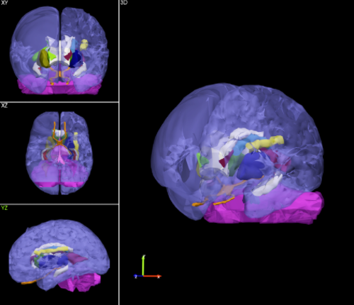
To obtain morphometric data about neurons or structures that extend beyond a single tissue section, creating a serial section reconstruction—a digital representation of aligned serial tissue sections—is extremely useful. You can reconstruct serial sections directly at the microscope or using images captured with Neurolucida.
Neurolucida includes tools that help you create neuron and neuroanatomical volume reconstructions quickly, including a tool for automatically tracing the outline of each tissue section or cytoarchitectural boundaries within the specimen. Once the reconstruction is complete, you can trace neurons and other structures of interest in each tissue section at a higher magnification and see the bigger picture with clarity.
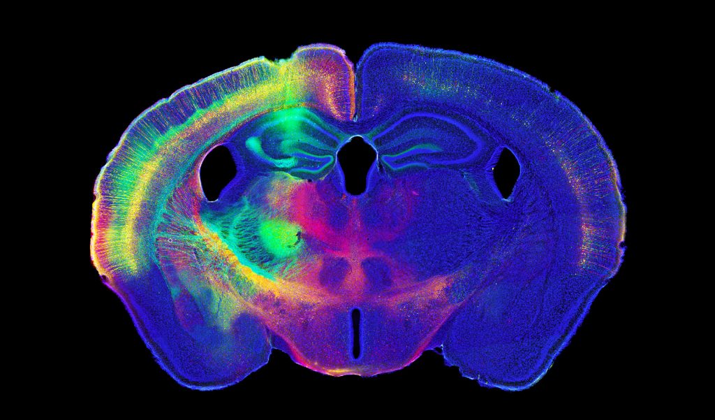
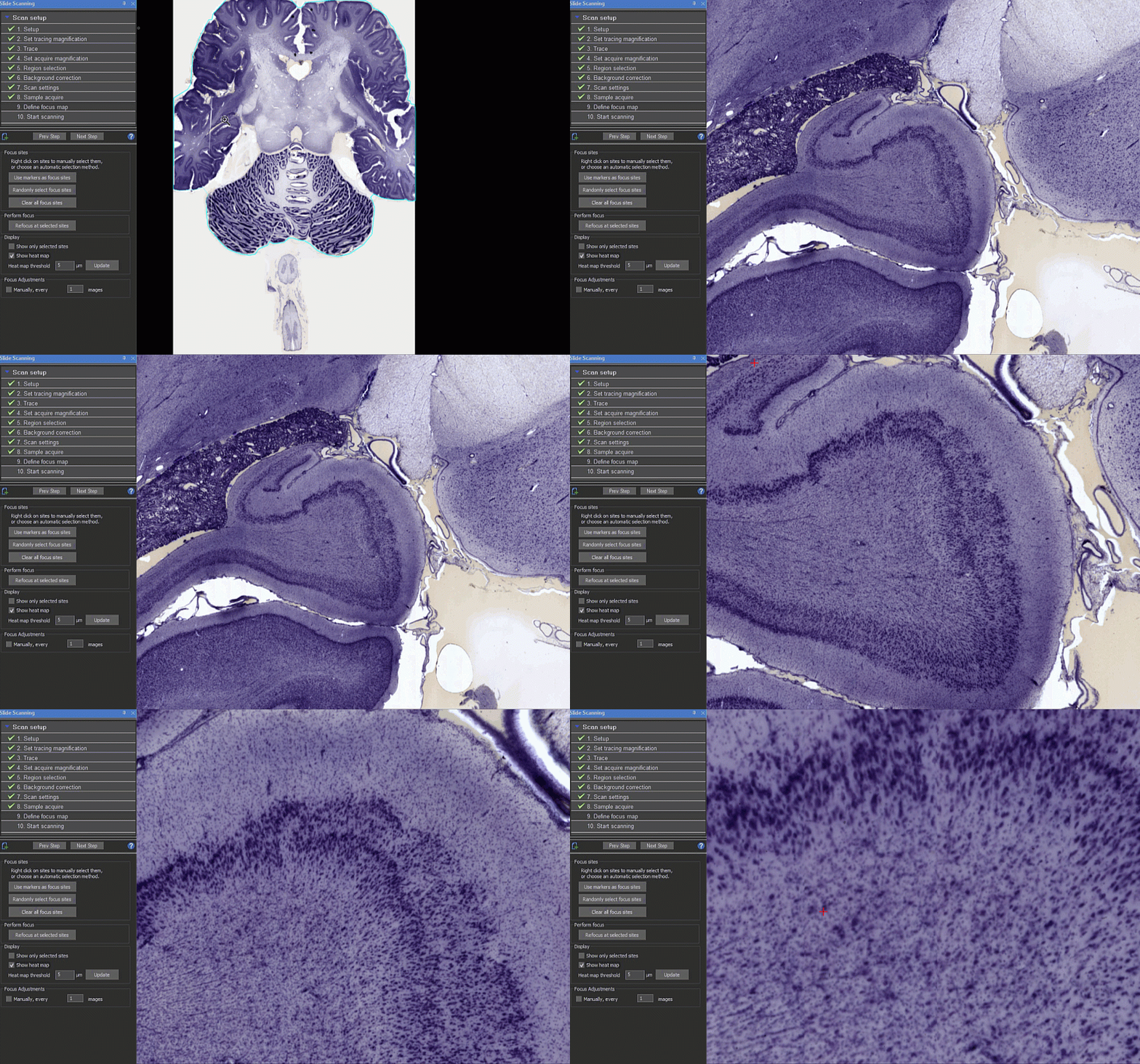
Neurolucida can serve as your microscope image acquisition system, too. With features to acquire high-quality images and z-stacks, your system can be used for much more than neuron tracing. You can also capture high-quality 2D and 3D whole-slide images (high resolution digital montages of your specimen) with the addition of the Slide-Scanning Module. Use image processing features such as background correction to insure clean, even, image quality images. Neurolucida can acquire a wide array of images, using brightfield and multi-channel fluorescence that surpass the quality of most slide scanners in a box.
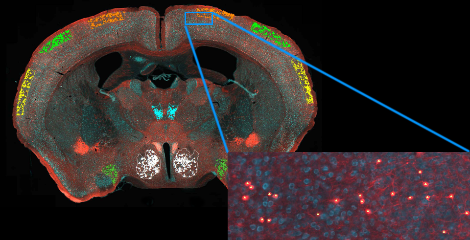
Identify labeled neurons in whole slide images using an intuitive cell detection workflow that incorporates advanced machine learning. Our deep-learning algorithms provide robust cell detection even when there are histologic and/or imaging artifacts such as edge effects, uneven illumination, and variable staining across specimens.
Neurolucida also supports importing image stacks and whole-slide images acquired using other systems. Working with whole-slide images enables you to create and analyze neuron tracings and reconstructions on any computer, even those not connected to a microscope.
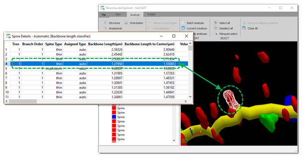
Neurolucida Explorer, the companion software included with Neurolucida, includes dozens of quantitative morphometric analyses that you can run on reconstructions and brain maps created using Neurolucida. Reconstruct and visualize your specimen with Neurolucida, then use Neurolucida Explorer to obtain quantitative data that extend your results. Developed in concert with neuroscientists using our products, Neurolucida Explorer can be used to analyze thousands of parameters. It includes an easy-to-use batch processing function and can export results that are compatible with other software such as Excel, Matlab, Python, and R.
Neurolucida is used across the globe by the most prestigious laboratories.


We are pleased to announce the release of Neurolucida®version 2024.1.3, which introduces key updates to offline software licensing. These updates

We are pleased to announce that the International Neuroinformatics Coordinating Facility (INCF) has endorsed the MBF Bioscience neuromorphological file format as

When we flex our thumb or point our finger, axons carry impulses from the brain to neurons in the spinal cord,
NeuroIucida’s utility is underscored by the number of references it receives in the worlds most important scientific publications.
Ament, S. A., M. Cortes-Gutierrez, et al.
A single-cell genomic atlas for maturation of the human cerebellum during early childhoodView Publication

Timonidis, N., M. Rubio-Teves, et al.
Analyzing Thalamocortical Tract-Tracing Experiments in a Common Reference SpaceView Publication

Jeon, K.-I., A. Kumar, et al.
Blocking Mitochondrial Pyruvate Transport Alters Corneal Myofibroblast Phenotype: A New Target for Treating FibrosisView Publication

Tetsushi, N., F. Satoshi, et al.
Microglia are dispensable for developmental dendrite pruning of mitral cells in miceView Publication

Qin, Y., X.-Y. Zhang, et al.
Downregulation of mGluR1-mediated signaling underlying autistic-like core symptoms in Shank1 P1812L-knock-in miceView Publication

Nevue, A. A., B. M. Zemel, et al.
Cell type specializations of the vocal-motor cortex in songbirdsView Publication

Hernandez-Reynoso, A. G., B. S. Sturgill, et al.
The effect of a Mn(III)tetrakis(4-benzoic acid) porphyrin (MnTBAP) coating on the chronic recording performance of planar silicon intracortical microelectrode arraysView Publication

Rasmussen, V. F., A. Schmeichel, et al.
Sweat gland nerve fiber density and association with sudomotor function, symptoms, and risk factors in adolescents with type 1 diabetesView Publication

Beers, D., D. Goniotaki, et al.
Barcodes distinguishing morphology of neuronal tauopathyView Publication
There are editions of Neurolucida available that work with, or without, a microscope. If the processes from your cells span multiple sections or dozens of fields of view, you may find it quicker and easier to reconstruct your cells with a microscope, using Neurolucida – Microscope Edition. If you’re reconstructing cells that are relatively easy to image, you may benefit from the automatic reconstruction features available in Neurolucida 360.
Neurolucida systems can be integrated with all 4 major microscope manufacturers (Zeiss, Olympus, Leica and Nikon). Depending on your microscope model, components, and experimental needs, your current microscope system may require hardware upgrades. Some of the most important components are stage, focus drive, cameras, and light sources. Contact us to assess your microscope system and help you determine a configuration that facilitates your research goals and fits your budget.
Neurolucida is ideal for reconstructing any cell or structure that has tree-like processes–many researchers use Neurolucida to reconstruct astrocytes, for example. Neurolucida also enables you to map and reconstruct anatomical regions through serial sections to create 3D models. You can acquire 3D image stacks and 2D or 3D scans of slides for publication or archival or image analysis away from the microscope.
The following MBF software modules require Neurolucida to function and can help improve your microscopic imaging process:
Every license of Neurolucida and Neurolucida 360 includes access to Neurolucida Explorer companion software. This data-analysis tool can quickly and easily export your data to Excel or to the clipboard. It even includes batch exporting so that you can process hundreds of data files with just a few clicks!
No, you will be given access to a free copy of Neurolucida Explorer with every copy of Neurolucida or Neurolucida 360 you purchase. If you don’t own Neurolucida or you need more licenses for use on analysis-only computers, you can purchase stand-alone Neurolucida Explorer licenses.
Neurolucida enables reconstruction of neurons directly from slides, and can connect to a wide range of microscopes, stages, and cameras. You can also import images and trace manually plane by plane in a 2D view. In contrast, Neurolucida 360 does not connect directly to microscope systems; it is designed for automatic, semi-automatic, and manual reconstruction of neuronal architecture from previously acquired images in a 3D environment. You can rotate, pan, and zoom while semi-automatically tracing or editing the computer-generated trace.
Neurolucida is ideal if your cells span across multiple sections or have distances across many fields of view, because reconstructing directly from the slide in live image mode tends to be faster than acquiring many images. If you’re working from images acquired on a confocal, 2-photon, or other microscope, we recommend Neurolucida 360.
Both Neurolucida and Neurolucida 360 support 3D serial section reconstructions, and offer modules for opening images and image stacks, MRI images, slide scans and other third-party formats.
"We are enjoying using your fantastic software and it is making a difference to our work."

Andrew Randall, Ph.D. University of Exeter Medical School
"Our system worked almost every day for the last eight years with no major failure (we published more than 15 papers using Neurolucida). A terrific machine!"

Ferdinando Rossi, M.D., Ph.D. University of Turin
"I have been using Neurolucida for over 20 years now. It sets the standard for what a tracing program should be able to do, and the ease of use has continually increased over the years. I have undergraduates using the program within a week. Moreover, the support MBF Bioscience provides is unmatched--they are the best."

Bob Jacobs, Ph.D. Colorado College
"We have been tracing dendrites for almost 30 years, using a variety of home-grown and commercial systems. We have used Neurolucida for almost 15 of those years now, and it is by far the most powerful, reliable, flexible, and easy to learn and use system of all that we’ve worked with. Neurolucida is clearly the product of people who thoroughly understand and appreciate quantitative neuroanatomy. This system has been absolutely indispensable to our research, resulting in over 25 publications. I can’t imagine our science without Neurolucida! Furthermore, the support provided by MBF has been simply outstanding, including program modifications, hardware support and development, troubleshooting, you name it - and always provided quickly, cheerfully, and knowledgeably. Nobody does it better!"

Dale Sengelaub, Ph.D. Indiana University
"I'm a huge fan Neurolucida's cell tracing and auto-detection features, which greatly assisted me in characterizing the morphology of abnormal neurons. Neurolucida is easily learned, intuitive, and was clearly designed by scientists."

Michael Hester, Ph.D. University of Cincinnati
"I have also been using Neurolucida for over 20 years with over 50 publications that have relied at least in part on the software. The product is marvelous and the support is the best of any software product I own. The work we do would be next to impossible without Neurolucida. The software is so intuitive that even high school students can do basic tracing almost immediately."

Ruth Stornetta, Ph.D. University of Virginia
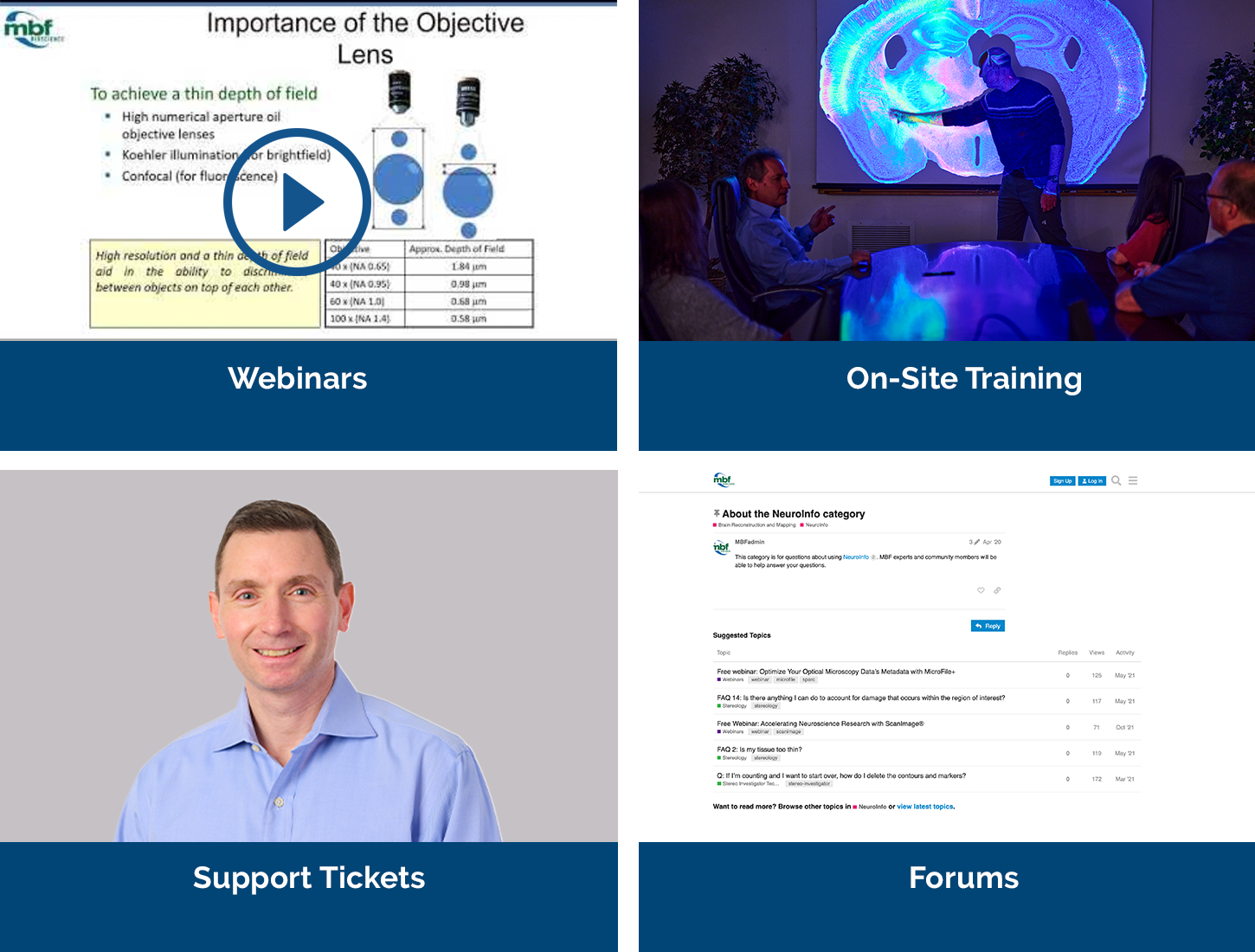
Our service sets us apart, with a team that includes Ph.D. neuroscientists, experts in microscopy, stereology, neuron tracing and reconstruction, and image processing. We’ve also developed a host of additional support services, including:
We offer a free expert demonstration of Neurolucida. During your demonstration you’ll also have the opportunity to talk to us about your hardware, software, or experimental design questions with our team of Ph.D. neuroscientists and experts in microscopy, neuron tracing, and image processing.


