
Stereo Investigator Software Update: Version 2024.1.3
We are pleased to announce the release of Stereo Investigator®version 2024.1.3, which introduces key updates to offline software licensing. These
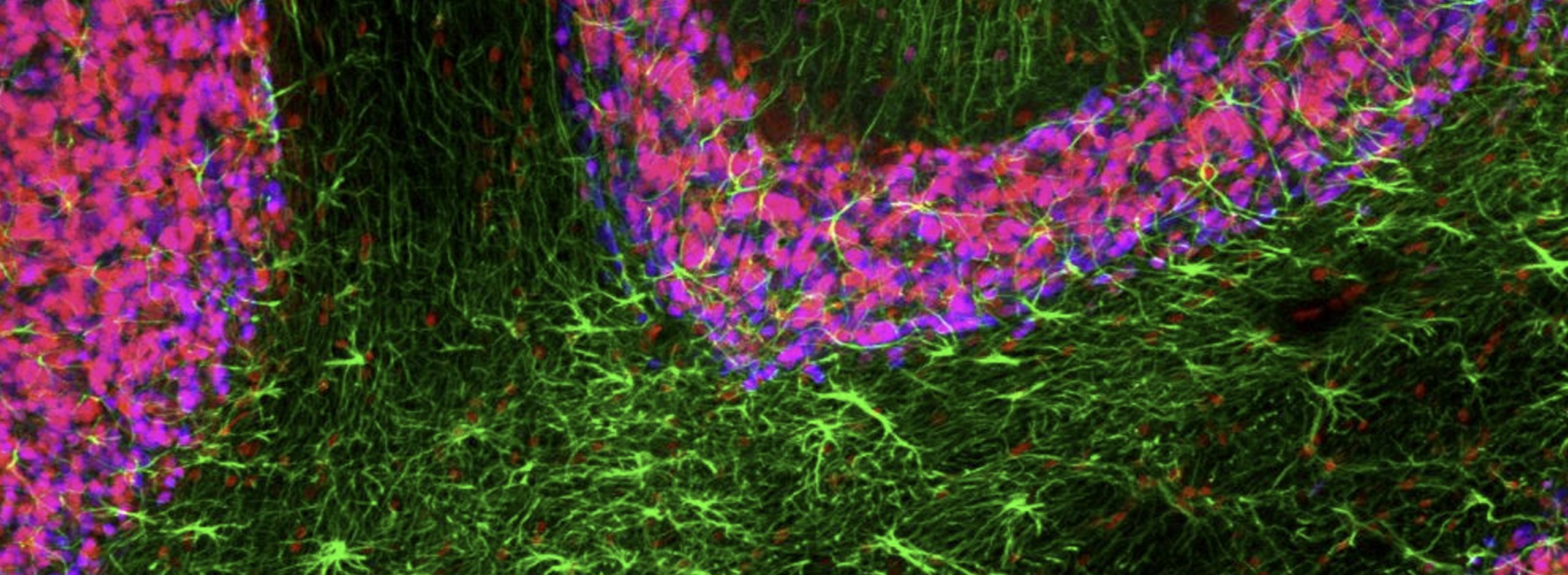
A Stereo Investigator system provides you with the means to make the most accurate, unbiased quantification of the number, length, area, and volume of cells, subcellular and macro structures in your tissue specimens. It is a key research tool that has helped scientists to make discoveries in numerous areas of neuroscience, including neurodegenerative diseases, addiction, autism, neuropathy, memory, and behavior, as well as other research fields including pulmonary and kidney research, and toxicology.
The Stereo Investigator system makes stereology accessible, with easy-to-follow workflows for the most commonly used probes and clear quantitative results. With Stereo Investigator, stereology can be performed directly on a microscope system, or from image stacks or whole slide images.
Visit our comprehensive stereology information site, stereology.info to learn more.
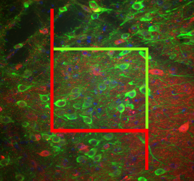
Stereology made accessible
With Stereo Investigator, you can perform unbiased quantification of cell populations, lengths of fibers or vessels, or area and volume of regions using design-based stereology, a set of methods for rigorous quantitative analysis of the size, shape, and number of objects. Stereo Investigator leverages unbiased statistical sampling, that helps enable researchers to count, measure, and precisely quantify visible features in microscope specimens using any of the 20+ included stereology probes without needing to examine the entire specimen to obtain statistically valid results. Stereo Investigator makes complicated stereological analyses simpler. It navigates you through the rigorous procedures required for unbiased quantitation or morphometric analyses and generates relevant calculations needed to generate final results and statistics. When needed or desired, you can also generate reviews and audits.
Stereo Investigator software includes sophisticated computerized microscope control that is powerful, flexible, and easy to use. In a complete Stereo Investigator system, which includes a microscope, computer, and Stereo Investigator software, all individual components work together harmoniously, setting the standard for state-of-the-art stereology systems.
Join the growing community of researchers who trust Stereo Investigator to help them obtain reliable results suitable for publication in high impact-factor journals over the last 20 years. With more than 8,000 published research papers, Stereo Investigator is cited 5+ times more frequently than its nearest competitor.
Stereo Investigator has been developed with support from the National Institute of Mental Health (NIMH)
| Minimum System Requirements | |
| Operating System | Windows 10, 64-bit |
| Processor | 4-core |
| CPU RAM | 16 GB |
| GPU RAM | >6 GB |
| Recommended System Requirements | |
| Operating System | Windows 10, 64-bit |
| Processor | 8-core |
| CPU RAM | 64 GB |
| GPU RAM | >8 GB |
| Storage | Solid state drive(s) |
| File Input Options | |
| Supported image file formats | CZI, HDF5, H5, IMS, JP2, JPG, JPEG, JPF, MJC, JPX, LIF, LSM, ND2, OIB, OIF, TIF, TIFF, SVS |
| Output Specifications | |
| Data output formats | XML*, ASC, DAT |
|
| * Our XML data file format, the Neuromorphological File Specification (NFS), is endorsed as a standard by the INCF. |
| Image file output formats | JP2, JPX, TIFF, SVG |
| Movie export format | MP4 |
| Hardware Compatibility | ||
|
Computer controlled microscopes | Leica supported microscopes:
| Zeiss supported microscopes:
Nikon supported microscopes
Olympus BX and IX Series:
|
| Manual microscopes | Most models after 2002 of Zeiss, Olympus, Nikon, Leica | |
|
Structured illumination |
| |
| Filter wheels and shutters | Zeiss, Olympus, Leica, Nikon, LEP, Prior, Sutter | |
|
Color video cameras |
| |
|
Monochrome video cameras |
| |
| Light sources |
| |
Supported Image File Formats: PDF
Contact us for an up-to-date list or question about specific models
Unprecedented study reveals structure-function adaptations in the facial nucleus of elephants
>> Learn More
Stereo Investigator System for Quantifying Neurotransmitter Switching in Human Brains Installed at Dr. Nicholas Spitzer’s Lab
>> Learn More
Differences Associated with Fetal Growth Restriction are Found Using Stereo Investigator
>> Learn More
Curcumin Lowers Neuroinflammation in Mouse Model
>> Learn More
MBF Bioscience research team contributes novel dendritic spine analysis in study published in Science
>> Learn More
Diet Restriction Slows Neurodegeneration and Extends Lifespan of DNA-Repair-Deficient Mice
>> Learn More
Uncovering the role of microglia in fetal alcohol spectrum disorders
>> Learn More
Stereological Study Reveals Neuron and Glia Proliferation in Hippocampus of Lithium-Treated Mice
>> Learn More
Iron Deficiency Worsens Fetal Alcohol Spectrum Disorders
>> Learn More
Genetic Mutation Accelerates CTE Pathology
>> Learn More
Dying neurons in Alzheimer’s patients show signs of improvement after gene therapy
>> Learn More
How Transplanted Stem Cells Behave in Injured Spinal Cord Tissue
>> Learn More
Delayed loss of neurons occurs in mice with mild TBI and anxiety
>> Learn More
Higher levels of pTau found in Alzheimer’s disease patients with psychosis
>> Learn More
New Neurons Erase Memories
>> Learn More
Scientists Use Stereo Investigator in Spinal Cord Injury Study
>> Learn More
Scientists Use Stereo Investigator in Spinal Cord Injury Study
>> Learn More
Scientists Map Photoreceptor Cells of Deep-Sea Sharks
>> Learn More
Humans Generate Most Cerebellar Granule Cells Postnatally
>> Learn More
Exercise Heals the Brain After Binge Drinking
>> Learn More
Scientists use Stereo Investigator to Discover that Part of the Hippocampus Shrinks in Socially Isolated Rodents
>> Learn More
Researchers from Quebec Delay Symptoms of Huntington’s Disease in Mouse Model
>> Learn More
Hawaii Scientists Measure Density of Parvalbumin-Interneurons With Stereo Investigator
>> Learn More
New Zealand Scientists Use Stereo Investigator to Develop a New Model for Human Extreme Prematurity
>> Learn More
Scientists Assess Kidney Damage with Stereo Investigator, Determine Ultrasound Prevents Organ Injury
>> Learn More
Munich Scientists Analyze Placenta Morphometry with Stereo Investigator
>> Learn More
UCLA Scientists Count Cells with Stereo Investigator in Study Identifying Compensating Regions in Brain Damage
>> Learn More
Increased Choline During Pregnancy Improves Learning in Down Syndrome Mice
>> Learn More
Study Links Increased Touch to Enhanced Neurogenesis in Adult Mice; Stereo Investigator Used for Quantification
>> Learn More
Wisconsin Scientists Use Stereo Investigator to Quantify Neurons Formed From Stem Cells
>> Learn More
Researchers Use Stereo Investigator to Identify Abnormalities in Autistic Brains
>> Learn More
Neurolucida & Stereo Investigator Help Uncover Cerebellar Granule Cells’ Role in Muscle Memory
>> Learn More
Stereo Investigator Contributes to Study Showing Low Zinc Levels Associated With More Cell Deaths in Spinal Cord Injury
>> Learn More
Stereo Investigator Helps Scientists Assess Damage in Rat Model of Ischemic Stroke
>> Learn More
Florida Researchers Study Traumatic Brain Injury With Stereo Investigator
>> Learn More
MBF Contributes to Recent PLOS Article on New Mouse Model for Huntington’s Disease
>> Learn More
Scientists at Duke Say Mice Have Features Associated With Vocal Learning
>> Learn More
Stereo Investigator Helps Harvard Scientists Study Social Isolation’s Effects on the Brain
>> Learn More
Yale Researchers Make Breakthrough in Possible Depression Treatment
>> Learn More
UVM Scientists Use Neurolucida and Stereo Investigator to Study Neurons in the Avian Iris
>> Learn More
John Hopkins University Scientists Quantify Neurons with Stereo Investigator
>> Learn More
UCLA Scientists use Stereo Investigator to Quantify Juvenile Neurogenesis in Mice
>> Learn More
DHA Supplementation Prior to Brain Injury May Reduce Severity
>> Learn More
Washington University Researchers use Stereo Investigator to Study Axonal Transport in Parkinson’s Model
>> Learn More
University of Kansas Researchers use Stereo Investigator to Map Fetal Brain Hypoxia Sites
>> Learn More
Stereo Investigator Assists Stanford Stroke Center Scientists in Stem Cell Research
>> Learn More
Toronto Scientists Get First Direct Measurement of Myometrial SMCs in Pregnant Rat Uterus With Stereo Investigator
>> Learn More
Scientists in Spain Track Recovery in the Auditory Cortex
>> Learn More
Stereo Investigator Helps Uncover Autonomic Diabetic Neuropathy Link
>> Learn More
Yale Scientists Say AAV5 is Best for Transduction in Primates
>> Learn More
Mount Sinai Scientists Research the Effect of Aging on Learning and Memory with Stereo Investigator
>> Learn More
Stereo Investigator Helps Washington D.C. Scientists Study the Emergence of Human Language
>> Learn More
Swiss Scientists Use Stereo Investigator to Study Schizophrenia
>> Learn More
Stereo Investigator Aids in Researching Familial Alzheimer’s Disease
>> Learn More
Stem Cell Transplants Aid in Spinal Recovery
>> Learn More
Download Stereo Investigator product sheet here.
Stereo Investigator® Version 2024.1.3
Released November 2024
Licensing Update
Update to offline software licensing. Stereo Investigator software requires reactivation on systems that do not have access to the internet. Instructions for offline software activation are included in the Stereo Investigator user guide.
View Full Version History Here.
The Optical Fractionator probe is often the most appropriate stereological tool for counting the number of cells (or other objects) within a 3D region of interest. It yields accurate, quantitative data using reliable, proven methodology. This seemingly basic knowledge about a specimen can be immensely important, for example, helping researchers determine whether an experimental treatment affects the total number of cells within a brain region when compared to an untreated group.
Stereo Investigator provides the Optical Fractionator workflow which offers an easy-to-use, easy-to-learn interface that walks users through the complex process of correctly using the Optical Fractionator probe. New lab members can learn the process and be up and running quickly, while avoiding counting mistakes. This probe relies on systematic random sampling to obtain a statistically valid and unbiased sample of the region of interest. Stereo Investigator overlays a 3D disector (as appropriate for the specimen) that indicates where in the specimen to count cells using the probe. Once counting is complete, results are automatically calculated and can be viewed within the software, exported to Excel, or copied to the clipboard.
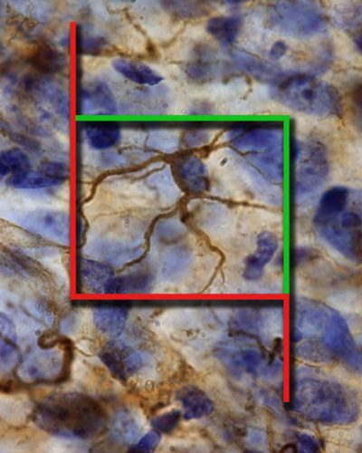
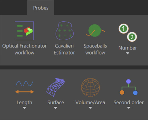
In addition to the widely used Optical Fractionator probe, Stereo Investigator includes over 20 other stereological probes and estimators to evaluate numbers of objects, length, surface area, or volume/area of regions of interest. If you are looking for morphometric data in your research, Stereo Investigator can help you collect it in a way that is unbiased and repeatable.
Some examples include estimating:
For fluorescence microscopy, where minimizing light exposure is typically a concern, you can conduct stereological analyses on acquired images, rather than on a live microscope-camera feed. If desired, Stereo Investigator can help you save time on the microscope and space on image-storage devices by acquiring images from a representative portion of your specimen using systematic random sampling (SRS). Once you’ve acquired a set of statistically representative image-stacks, you can run many stereology probes for different types of morphometric analyses and never have to worry that your fluorescence signal will fade. In addition, you can repeat analyses easily if desired.
For SRS image/image-stack acquisition, Stereo Investigator directs the motorized stage, camera, and light sources. You can configure image-acquisition parameters, then let Stereo Investigator automatically capture image stacks at each SRS site. Perform stereological analysis on the acquired images using Stereo Investigator installed either on the microscope-control computer or on a different computer with the Desktop version of Stereo Investigator software.
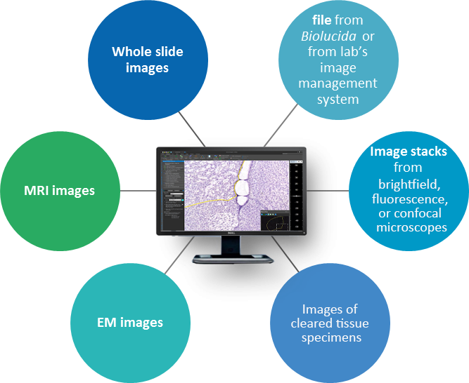
| Feature Description | Stereo Investigator MICROSCOPE EDITION | Stereo Investigator DESKTOP EDITION | ||
|---|---|---|---|---|
| The gold standard for unbiased stereology | ||||
| Quantifies number, length, volume, & surface area of cells, structures, & regions | ||||
| Works with 3D images | ||||
| Analyzes large 3D images aquired from light sheet and confocal microscopes | ||||
| Works with 2D images | ||||
| Analysis with images acquired from slide scanners | ||||
| Integrated microscope control to work directly with live images at the microscope | ||||
| Acquire 3D/2D images for Systematic Random Sampling (SRS) |
Stereo Investigator is used across the globe by the most prestigious laboratories.


We are pleased to announce the release of Stereo Investigator®version 2024.1.3, which introduces key updates to offline software licensing. These

Using specimens that were collected over three decades from zoos, researchers at Humboldt University of Berlin examined facial motor control in

We are pleased to announce that the International Neuroinformatics Coordinating Facility (INCF) has endorsed the MBF Bioscience neuromorphological file format as
Stereo Investigator has been cited in over 8,000 published research papers. 5 times more than its nearest competitor.
Rentsch, P., T. Egan, et al.
The ratio of M1 to M2 microglia in the striatum determines the severity of L-Dopa-induced dyskinesiasView Publication

González-Salvatierra, S., C. García-Fontana, et al.
Cardioprotective function of sclerostin by reducing calcium deposition, proliferation, and apoptosis in human vascular smooth muscle cellsView Publication

Bautista, J., M. Á. García-Cabezas, et al.
Pattern of ventral temporal lobe interconnections in rhesus macaquesView Publication

Wang, Z., D. Zheng, et al.
Enabling Survival of Transplanted Neural Precursor Cells in the Ischemic BrainView Publication

Villar-Conde, S., V. Astillero-Lopez, et al.
Synaptic involvement of the human amygdala in Parkinson’s diseaseView Publication

Stimpson, C. D., J. B. Smaers, et al.
Evolutionary scaling and cognitive correlates of primate frontal cortex microstructureView Publication

Most Stereo Investigator systems are microscope based. It’s often fastest and easiest to evaluate specimens directly on the microscope. We also offer the Stereo Investigator Desktop Edition that does not include microscope-control elements. With the desktop edition, you simply open previously acquired 2D images or 3D image stacks and perform the probe away from the microscope, freeing up the microscope for other uses.
Stereo Investigator can integrate with all 4 major microscope manufacturers (Zeiss, Olympus, Leica, and Nikon), but depending on your microscope model, components, and experimental needs, your current microscope configuration might require upgrades or selective substitution of components. Some of the most critical components are stage, focus drive, cameras, and light sources. Please contact us to assess the compatibility of your existing microscope hardware and help you configure the optimal Stereo Investigator system for your research.
The following MBF software modules require Stereo Investigator to function and can help improve your microscopic imaging process:
Stereo Investigator: Microscope Edition is the most versatile and comprehensive version. It enables you to perform stereology directly on a microscope by controlling your microscope components, such as the motorized stage and camera. In addition to running nearly all stereological probes, you can document your experiments by acquiring images with sample and region localization so that it is easier to navigate and record your experiment.
Stereo Investigator: Desktop Edition has all the same probes as Microscope Edition, but is intended for use away from the microscope. With this version, you can work from 2D images or 3D image stacks acquired previously. This frees up busy microscopes for other users and uses.
Yes! In addition to stereological analysis, Stereo Investigator offers the 3D Serial Section Reconstruction module. This enables you to map and reconstruct your regions of interest, view your data in 3D, and create and export movies or models for presentations.
"Our Stereo Investigator system works perfectly. We use it every day and don't have any problems whatsoever. Thanks for the good system."

Grazyna Rajkowska, Ph.D. The University of Mississippi Medical Center
"I have been using the Neurolucida and Stereo Investigator software from MicroBrightField for the last 5 years, and this software has become an essential part of my research program. Whether it is 3 dimensional reconstruction, counting particular particles or mapping the developing and adult vascular, these programs are a scientific must. I personally have been able to push my research in ways that would otherwise not have been possible. Furthermore, the ability to quantitate and detect subtle differences is and will continue to be critical to my scientific program. I would highly recommend these companion programs to any research scientist."

Sunder Sims-Lucas, Ph.D. Nationwide Children's Hospital
"If there is another program for unbiased stereology, I don't know what it is. This is the gold standard, and I can't say enough to praise the technical support provided by MBF Bioscience."

Bob Jacobs, Ph.D. Colorado College
"We've used Stereo Investigator for 10+ years. The technical support is outstanding, and the company itself it highly responsive to their customers."

Glenn Rosen, Ph.D. Beth Israel Deaconess Medical Center
"We have used Stereo Investigator for over 10 years now. The service and technical support is always great and they keep adding new updates with more tools. "

Paula Bickford, Ph.D. University of South Florida
"Stereo Investigator is a excellent stereology system. It is easy to work with and the workflows help you to work very efficiently. It is a crucial research tool for obtaining estimates of number of neurons, length of capillaires, length density of fibers, volume measurements of regions of interests or neurons, etc in specific brain regions. I would like to recommend it to everybody who is interested in stereology. In addition, MBF provides excellent technical support and helps you to find the best technical tools for your research challenges on morphometry."

Wilma Van De Berg, Ph.D. VU University Medical Center - Neuroscience Campus Amsterdam
"Stereo Investigator is an excellent research tool for unbiased stereology. In our research group when we set out to quantify the complex network nerves residing in the stomach mucosa. With Stereo Investigator we were able to get accurate estimates of numbers and show differences between your study groups without the introduction of sampling bias."

Mona Selim, Research Associate University of Minnesota
"Stereo Investigator has been the workhorse of our Microvascular Pathology Lab for the last six years. We have been able to measure vascular density with an accuracy never before available to our studies and more recently SI has provided efficient and precise comparisons of hypoxia and ischemic damage in cerebral vessels and neurons. The helpfulness, availability and experience of the MBF staff has been extraordinary. I would recommend any of their products wholeheartedly."

Clara Thore, Ph.D. Wake Forest University
"Stereo Investigator is great for stereology. Although there is no field representation in the UK, the team in Vermont have been excellent at supporting us in our research applications. With online support and real time meetings and webinars, we've been able to make substantial progress on our research projects. MBF aren't afraid of a technical challenge, which is excellent, and have managed to provide us an excellent solution for our complex research needs."

Ann Wheeler, Advanced Imaging Facility Manager Queen Mary, University of London
"Stereo Investigator was particularly useful for estimating cell density in larger brain structures, such as the inferior colliculus."

Matt Pitts, Ph.D. University of Hawaii
"Since we purchased our system four years ago, our experience with the MBF equipment and especially the MBF people has been outstanding. I cannot speak any higher about their professionalism and attention for our needs. "

Bogdan A. Stoica, M.D. University of Maryland, School of Medicine
"Stereo Investigator is powerful yet easy to use. The staff at MBF Bioscience is always helpful answering our questions and recommending optimal ways to conduct our stereological analyses."

Matthew Goldberg, Ph.D. University of Texas Southwestern Medical Center
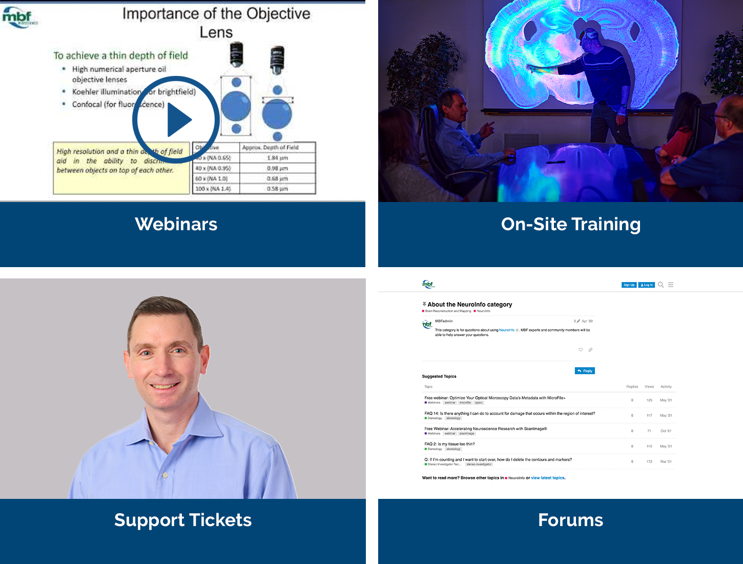
Our service sets us apart, with a team that includes Ph.D. neuroscientists, experts in microscopy, stereology, neuron reconstruction, and image processing. We’ve also developed a host of additional support services, including:
We offer a free expert demonstration of Stereo Investigator. During your demonstration you’ll also have the opportunity to talk to us about your hardware, software, or experimental design questions with our team of Ph.D. neuroscientists and experts in microscopy, neuron tracing, and image processing.


