First System for Imaging and Analyzing Large Mammalian Brain Tissue Installed in South Africa

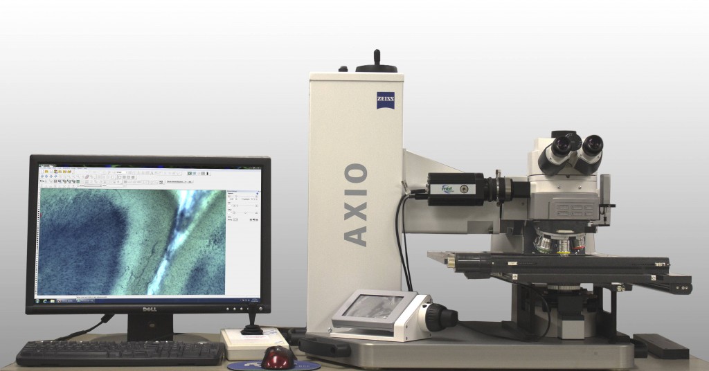
Dr. Paul Manger’s new system developed by MBF Bioscience for analyzing large mammalian brain specimens
Dr. Paul Manger, Research Professor at the University of the Witwatersrand, South Africa, studies the brain anatomy of various animals to better understand the relationship between brain structure and function. Some of the mammals he studies have very large brains that don’t fit under a conventional research microscope. A team of scientists and microscopy experts at MBF Bioscience developed a new microscope system that enables Dr. Manger to analyze and image whole sections of large brain tissue. This system is the first for imaging and analyzing entire tissue sections of large mammalian brains. It will give Dr. Manger unprecedented analyses of African mammal brains and a way to share large images of tissue specimens with collaborators or a global audience.
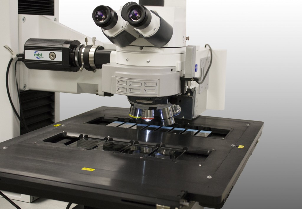
To accommodate the large tissue specimens, MBF Bioscience developed the system with a Zeiss Axio Imager Vario microscope which is used in the semiconductor industry for inspecting silicon wafers. The motorized stage, built by Ludl, is enormous – it can hold tissue specimens with dimensions of up to 30cm x 40cm. This hardware, combined with Neurolucida, Stereo Investigator, and Biolucida software, allows Dr. Manger to capture 2D and 3D whole slide images of large tissue specimens, quantitatively analyze the specimens, and share the images with other scientists in a repository of African vertebrate brains. This type of imaging and analysis of large brains is not currently done in any other laboratory.
Dr. Manger now has the tools to help him uncover new information about the sensory, motor and cognitive capacities of African mammals. And, he has a new way to disseminate that information to scientists and collaborators who can build upon the findings to further the understanding of mammalian brains.
A sample of Dr. Manger’s publications can be found below.
Quantitative analysis of neocortical gyrencephaly in African elephants (Loxodonta africana) and six species of cetaceans: Comparison with other mammals
Organization and number of orexinergic neurons in the hypothalamus of two species of Cetartiodactyla: A comparison of giraffe (Giraffa camelopardalis) and harbour porpoise (Phocoena phocoena)
Neuronal morphology in the African elephant (Loxodonta africana) neocortex


