Scientists in Portugal Use Neurolucida Explorer to Analyze Neuroplasticity in Depression
Life’s little pleasures often elude those suffering from depression, including rats, who show little interest in sugar water after experiencing stress. This behavior leads scientists to speculate that the illness might be characterized by a defect in the brain’s neural reward circuit.
Recent research focuses on a key element of this circuit – the nucleus accumbens (NAc), part of the brain region known as the ventral striatum, which is thought to regulate motivation and reward processing. In a new study of stress-induced depression in rats, researchers at the University of Minho in Braga, Portugal saw morphological changes in the dendrites of medium spiny neurons in the NAc, alongside disturbances in gene expression in this region. They also saw these changes reversed after administering antidepressants.
By using Neurolucida Explorer to analyze 3D reconstructions of medium spiny neurons generated with Neurolucida, the researchers observed longer than normal dendrites and greater spine density in the depressed rats. According to the paper, these findings contrast with studies of the hippocampus and prefrontal cortex, where chronic stress leads to shorter dendrites.

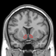
Nucleus Accumbens
The authors say the morphological changes are related to changes in gene expression, including increased brain-derived neurotrophic factor (BDNF) expression and other genes related to neuroplasticity.
“These observations add to the evidence that neuroplastic changes in the NAc contribute to the pathophysiology of depression and its pharmacologically-induced recovery, and point to the role of the NAc in the regulation of (an)hedonia [diminished interest or pleasure],” the authors say in their paper published last month in Translational Psychiatry.
To see how stress-induced depression affected cell volume in the NAc, the research team conducted a stereological analysis of the region. But after quantifying neurons with Stereo Investigator, they did not see significant differences in the number of neurons in depressed rats versus controls.
“Given that this region is remarkably interconnected with the hippocampus and the prefrontal cortex, it remains to be demonstrated whether the changes herein described are causal or a mere consequence of the neurodegenerative effects triggered by stress in these ‘cortical’ regions,” the authors say.
Bessa, J., Morais, M., Marques, F., Pinto, L., Palha, J., Almeida, O., & Sousa, N. (2013). Stress-induced anhedonia is associated with hypertrophy of medium spiny neurons of the nucleus accumbens. Translational Psychiatry, 3(6), e266. doi:10.1038/tp.2013.39


