SVI Partnership Means Clearer Images for Researchers Worldwide
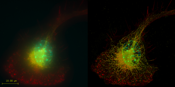
The microscopic world just got a whole lot clearer for scientists around the world who use MBF Bioscience software. When images are magnified as intensely as they are in today’s world of highly advanced scientific research, they don’t always appear crystal clear. But by incorporating the clarifying process of deconvolution to microscopic images, previously hidden details emerge, allowing scientists to work with greater facility.
Deconvolution is a processing technique that improves the appearance of microscopic images by removing blurriness.
Thanks to our new partnership with the Dutch company SVI (Scientific Volume Imaging, BV), researchers who use Neurolucida and Stereo Investigator will see images with greater clarity than ever before.
“We’re always striving to find ways to help scientists become more productive and to open new pathways to discovery,” said MBF Bioscience founder and CEO Jack Glaser. “We’ve incorporated SVI’s Huygens technology into our own software, Neurolucida and Stereo Investigator, and we’re excited by this opportunity to make SVI’s Huygens deconvolution software available to scientists doing other kinds of research as well.”
{Image: Macrophage fluorescently stained for tubulin (yellow), actin (red) and the nucleus (DAPI, blue). Left: original image, recorded with a wide field microscope. Right: the same dataset, deconvolved using Huygens Professional. Data courtesy of Dr. James Evans, Whitehead Institute, MIT Boston MA, USA. Image courtesy of SVI}


