Stem Cell Transplants Aid in Spinal Recovery
Thousands of people in the United States have spinal cord injuries (SCIs), with associated loss of movement and sensation below the site of the injury. Neural and glial cell transplants into research animals after SCI have correlated with recovery of function. The improvement may be caused by the transplanted cells; it’s thought that remyelination by the transplanted glial cells is the main reason for the improvement. Also, if adult neural stem cells are transplanted, there is evidence they form new neurons. In “Analysis of Host-Mediated Repair Mechanisms after Human CNS-Stem Cell Transplantation for Spinal Cord Injury: Correlation of Engraftment with Recovery” (2009, Hooshmand MJ, Sontag CJ, Uchida N, Tamaki S, Anderson AJ, Cummings BJ, PLoS One) the authors use Stereo Investigator’s powerful quantitative tools to determine whether changes in the host environment may also be correlated with improved function.
Using Stereo Investigator, specific aspects of the host milieu were compared between spinal cord injured animals (Non-Obese- Diabetic-severe combined immunodeficient mice) that received transplants of human central nervous system-stem cell neurospheres (hCNS-SCns) and those that did not. The sampling parameters such as section interval, grid size, and counting frame size, were determined by checking the coefficient of error to make sure it was low. In some cases, additional post-hoc power analysis of data from previous publications was used to demonstrate that the parameters were appropriate for the required precision. Serotonergic fiber length was estimated using the Isotropic Virtual Planes probe with a 60X objective. Blood vessel length was estimated using the Space Balls probe with a 40X objective. The areas and volumes of lesions, spared tissue, and astrogliosis, were estimated using the Cavalieri probe with a 20X objective. Stereological results were complimented by biochemical protein analysis. In addition, the Optical Fractionator probe was used to estimate a non-host parameter, the number of neurons that live and proliferate from the hCNS-SCns transplant.
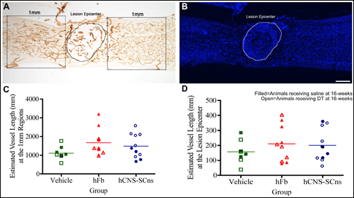
There were no differences found in the host characteristics between hCNS-SCns transplant animals and control animals. For example, there was no difference in the length of blood vessels. Platelet/ endothelial cell adhesion molecule immunohistochemistry was used to identify blood vessels. Some treatments following CNS trauma may promote behavioral recovery associated with vascular remodeling. Blood vessel length was estimated at the injury center, one mm rostral, and one mm caudal to the injury. There was no statistical difference between controls and hCNS-SCns transplanted animals (see figure, controls are vehicle and human fibroblasts (hFb)). Regarding the non-host characteristic of how many transplanted cells lived, multiplied, and migrated, the Optical Fractionator estimate showed that the transplanted cell number increased 194 percent after transplantation and migrated from the injection site. Ablation of some transplanted cells with Diptheria toxin correlated with a loss of locomotor recovery. This study shows that the direct consequences of the transplanted cells such as proliferation - correlate with improved function – while the transplant does not have an effect on host characteristics such as lesion volume, spared tissue, fiber sprouting, and angiogenesis, ruling out any correlation of an indirect effect of the transplanted stem cells with recovery.
Dan Peruzzi is a staff scientist at MBF Bioscience.
First published in The Scope, fall 2009.


