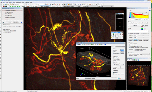
MBF Unveils Neurolucida and Stereo Investigator 9
Image courtesy of Juan Carlos Tapia, PhD, Harvard University
Now available, release 9 of our flagship Neurolucida and Stereo Investigator systems represent one of the largest changes in the history of these products. With many new features and an improved interface, we’re listening to you, our loyal customers, and delivering products with innovation, power, and speed.
Stereo Investigator and Neurolucida New Features
Image Handling: We’ve been busy when it comes to handling and visualizing your valuable image data. High bit depth, Nchannel (up to 16 bits per channel) image files are now accessible via an intuitive user interface. Acquire up to 6 independent channels of high bit depth data per image from any of our cameras. You can even load dozens of image stacks at once. Our new image handling lets you work with the latest image file formats from the major confocal microscope manufacturers. We’ve enhanced your ability to work with virtual slides to include 3D virtual slide stacks and allow you to save portions of these large montage images (and stacks) to separate files. All of this is controlled by a new image display control feature providing precise histogram adjustments.
Automatic Contouring: Automatically trace contours while you watch the software identify the tissue boundaries from images, image stacks, virtual slices, and even off the live camera feed. If you need to move the stage, the software does it automatically and continues tracing the outline of your region.
Automatic Object Detection: Now with just a few clicks you can let the software find cells, synapses, grains, or similar objects of interest in your images. Mark and count the objects or ask our software to automatically trace around the outside of an object. JPEG2000 Image File
Support: We now offer state-of-the-art image file storage with JPEG2000 support, which dramatically reduces disk space usage for all your image data. For instance, you’ll see image stacks that were 100 MB shrink to about 5 MB, depending on the image data and compression ratio you choose. Create very large virtual slices that exceed the computer’s physical memory by using JPEG2000 support, and store these files
using manageable file sizes. An example, we recently captured a large virtual slice (556,800 x 520,000) that compressed to only 40 GB; with earlier technology, it saved as an 800 GB image file.
Multiple User Setup: If you have a system that serves many users, you’re life just got easier. Use one login with all users or give each user a private login with their own customized settings. If you have a system with multiple hardware setups, this also makes it easy to keep the settings separate and switch between them.
Support for Aperio and Hamamatsu Nanozoomer Files: Now you can load Aperio SVS and Nanozoomer virtual slide files into Neurolucida or Stereo Investigator for analysis.
Improved 3D Visualization: A fully redesigned 3D visualization window makes it easier to view, manipulate, and understand image stacks and tracings. We’ve included cut planes in the X, Y, and Z axis from any view angle. Manipulate the image transparency to get the look you want from your image and tracing data. Look for the new automatic rotation tool that lets you adjust the angle of rotation
and speed.
Hardware: We’ve added support for monochrome and color cameras under 64-bit Vista and Windows 7. Our streamlined camera interface gives you the image you desire easily and quickly. Now, swap between multiple cameras without exiting the application to easily utilize the multiple camera ports on your microscope. Zeiss AxioCam, QImaging, and Baumer cameras are part of our line as well, along with tight integration for the Zeiss ApoTome and support for the Qioptic Optigrid structured illumination offerings.
Simplified Menus and Hot Keys: Streamlined workflows with more intuitive menus and dialog, along with new navigation keys help you maneuver around with less effort.
Drag and Drop: Drag and drop your images, image stacks, and data files for easier loading and display.
Tracing with Transparency: Ever want to look at or trace something where the tracing was in the way? Now simply adjust the transparency of the tracing to see the underlying image while tracing.
Serial Sections: Do you trace your sections separately and combine them later? We’ve made it much easier and intuitive to combine files together into a complete data file with fewer steps. Use the improved Serial Section Manager to define many sections in one step and view multiple sections.
These are just some of the new enhancements found in version 9.
More Neurolucida 9 Additions
Neurolucida has added new analysis tools for tracing and classifying dendritic spines, and improved editing features to correct your AutoNeuron tracings.
And More In Stereo Investigator 9
Stereo Investigator has a number of other new features including the Connectivity Assay (Pulmonary Edition). New enhancements for the Optical Fractionator include faster counting of multiple cell populations, new displays for the results, and improved Excel export for easier and more comprehensive reports. The Area Fraction Fractionator is also faster and more streamlined. An important
new tool, the Oversample and Resample allows you to methodologically pick the most efficient sampling parameters for your material.
First published in The Scope, fall 2009.
If you enjoyed this article, fan us on Facebook and follow us on Twitter to get the latest updates on MBF Bioscience company and customer news.


