Whole Brain Analysis

MBF Bioscience > Whole Brain Analysis

Revolutionizing Whole-Brain Analysis


Introduction


Understanding the intricate mechanisms underlying brain function and behavior remains one of the most complex challenges in modern neuroscience. Unraveling these mysteries necessitates a comprehensive, multi-dimensional approach that integrates diverse data streams, including neuronal activity patterns, anatomical connectivity, cellular morphology, and gene expression profiles across the entire brain[1]. However, compiling and visualizing such vast, heterogeneous datasets presents significant challenges, hindering our ability to derive meaningful insights.

Current Challenges in Whole-Brain Analysis


The recent explosion in molecular neuroanatomical tools is revealing the complexity of neural circuits underlying behavior in unprecedented ways. Applying these tools has provided comprehensive multi-dimensional databases of single neuron gene expression profiles, neuroanatomical connectivity, cellular morphology, and neuronal activity patterns mapped into common reference atlases. Evident from the wealth of information in these data is the necessity to incorporate a multi-dimensional, whole-brain approach for understanding specific linkages between brain circuits and normal behaviors or disorders.


However, existing software solutions often struggle to handle the large-scale, multi-dimensional data generated by diverse neuroanatomical techniques. Many tools are limited to specific brain atlases, model organisms, or data types, constraining researchers’ ability to synthesize and explore data holistically[2].

The NeuroInfo Solution


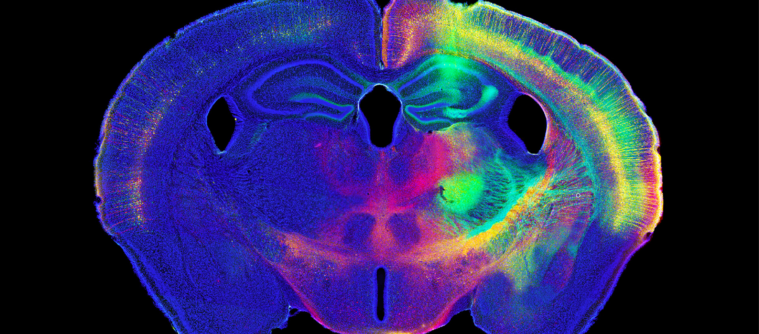
In response to these challenges, we have developed NeuroInfo, an integrated software application designed to revolutionize whole-brain analysis. Leveraging cutting-edge deep learning and registration technologies, NeuroInfo empowers researchers with comprehensive tools for labeling and analyzing the connectional architecture of neural circuits across the entire brain.


NeuroInfo can be used with both serial sections imaged with fluorescence microscopy and cleared whole brains imaged with light sheet microscopy, offering advantages for each method:


  • Serial sections can be processed with a wide range of immunohistochemical approaches and imaged with high-magnification objective lenses, providing access to labeling methods not amenable for use on cleared whole brains.
  • Cleared whole brains offer the benefit of analyzing the entire brain without artifacts from tissue sectioning, although some labeling techniques may not be compatible with this approach.

Key Features and Innovations of NeuroInfo


1. Deep Neural Networks:
NeuroInfo‘s deep neural networks are trained on meticulously annotated data, enabling accurate identification and mapping of labeled cells while accounting for local variances in staining patterns. The algorithms are optimized for precise cell detection and quantification, even in the presence of uneven staining or low target protein abundance [3].


2. Automated Atlas Registration:
NeuroInfo seamlessly aligns the detected cells to widely accepted brain atlases like the Allen Mouse Brain Atlas and Waxholm Rat Atlas, facilitating comprehensive whole-brain analysis. The automated registration process ensures that findings can be contextualized within a standardized anatomical framework, enabling comparisons across experiments and laboratories[4].


3. Multi-dimensional Data Visualization:
NeuroInfo’s powerful visualization engine enables the creation of intricate 3D renderings that merge anatomically aligned data from diverse sources, including neural activity, connectivity, morphology, and gene expression profiles[5]. Researchers can explore and integrate these multi-dimensional datasets within a unified and interactive environment.


4. Robust Quantitative Analysis:
MBF Bioscience has a long track record of providing neuroscience researchers with sophisticated quantitative analysis tools. NeuroInfo extends this expertise by offering measurements such as the numbers, sizes, locations, and intensities of cells and important structures like injection sites.


Benefits for the Neuroscience Community


NeuroInfo streamlines the exploration of whole-brain circuits, neuronal populations, and biochemical marker expression by leveraging widely-used brain atlases as anatomical references. The software supports a wide range of brain imaging techniques, making it a versatile tool for laboratories using different approaches.


Advanced AI segmentation and registration methods ensure accurate and repeatable results, while user-friendly workflows make these powerful tools accessible to researchers without extensive programming expertise. This democratization of advanced analysis capabilities is crucial for advancing neuroscience research[6].

Key benefits of NeuroInfo include:


  • Automatic alignment of 2D and 3D images to standardized brain atlases
  • Brain-wide detection of cells and biochemical markers using state-of-the-art deep learning approaches
  • Compatibility with various brain imaging modalities, from whole-slide scanners to light sheet microscopy
  • Standardized and repeatable data generation for rigorous comparisons across studies and laboratories
  • User-friendly workflows that empower researchers without requiring programming expertise
  • Potential to foster interdisciplinary collaboration and accelerate neuroscience discoveries

Conclusion


NeuroInfo represents a paradigm shift in whole-brain analysis, empowering researchers with cutting-edge tools to integrate and interpret the vast, complex datasets essential for unraveling the neural underpinnings of behavior. By leveraging advanced deep learning methods, virtual reality annotation, and standardized brain atlases, NeuroInfo enables brain-wide characterization of cell populations and biochemical marker expression with unprecedented accuracy and efficiency.


Through its intuitive and interactive visualization capabilities, NeuroInfo transcends the limitations of traditional software solutions, fostering interdisciplinary collaboration and facilitating the synthesis of multi-dimensional data streams. As neuroscience continues its quest to unravel the intricate workings of the brain, NeuroInfo stands poised to catalyze groundbreaking discoveries and propel our understanding of brain function to new frontiers.

Fields Of Study

MBF Bioscience products are used for whole brain analysis in numerous ways, including the following:

Our Solutions for Whole Brain Analysis

BrainMaker®

BrainMaker automatically creates full-resolution, 3D reconstructions of the entire brain (or any organ) from serial sections of whole slide images.


NeuroInfo®

NeuroInfo enables whole brain experimentation for laboratories of every size. Neuronal populations and circuits are automatically characterized in brain volumes and overlain with brain region delineations in a standardized reference space to provide visual and quantitative anatomic context.


Vesselucida® 360

Vesselucida 360 is designed to quickly segment and fully reconstruct vessels and microvasculature in 3D to obtain reliable data about the length, connections, and complexity of vessels and microvessels.


Neurolucida® 360

Neurolucida 360 is the premier tool used by neuroscientists to quickly and accurately reconstruct intricate neuronal structures that range in scale from complex, multicellular networks of neurons to sub-cellular dendritic spines and putative synapses.

Want Help Understanding Which Product is Right for Your Application?

At MBF, we’ve spent decades understanding the needs of researchers and their labs — and have a suite of products and solutions that have been specifically designed for the needs of today’s most important and advanced labs. Our commitment to you is to spend time with you discussing the needs of your lab — so that we can make sure the solutions we provide for you are exactly what you’ll need. It’s part of our commitment to supporting you — before, during, and after you’ve made your decision. We look forward to talking with you!

References


[1] Bhatia et al. (2021). Integrated multi-scale brain atlases: A powerful tool for understanding brain function and disorders. Trends in Neurosciences.

[2] Verge et al. (2022). Limitations of thresholding methods in quantifying neuronal activity markers. Nature Methods.

[3] Shen et al. (2017). Deep learning for neuroimaging: a review. Neurocomputing.

[4] Niedworok et al. (2016). aMAP: a validated pipeline for registration and segmentation of brain images in R. Nature Protocols.

[5] Niedworok et al. (2019). Integrating brain-wide connectivity and gene expression to study neurodegenerative diseases. Brain Connectivity.

[6] Yuste et al. (2020). A community-based transcriptomics classification and nomenclature of neocortical cell types. Nature Neuroscience.

[7] Kim et al. (2021). Advances in whole-brain mapping: From micro to macro scales. Frontiers in Neuroanatomy.