* Please note pricing may vary outside the US
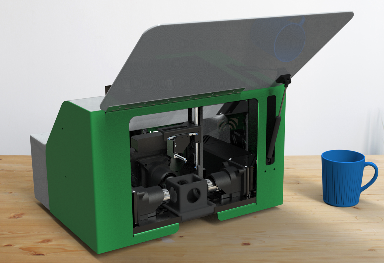
SLICE™- Revolutionary Light-Sheet Microscopy System
Product Overview
SLICE: a paradigm shift in light-sheet microscopy that combines high performance with unprecedented affordability and a compact device footprint. Invented by leading researchers at Columbia University, SLICE delivers advanced 3D imaging capabilities to research labs of all sizes. This innovative system offers high-resolution imaging of biological samples, making it ideal for both individual laboratories and core facilities. SLICE comes equipped with powerful software for giga-voxel visualization, seamless image stitching, and sophisticated post-processing. Moreover, it seamlessly integrates with MBF Bioscience’s renowned suite of analytical tools, including Neurolucida 360, NeuroInfo, and NeuroDeblur, providing a comprehensive solution for cutting-edge research in neuroscience and beyond.
Complete SLICE light sheet systems for less than $75k USD*

Key Benefits
- Affordable. Performance on par with systems costing ten times as much
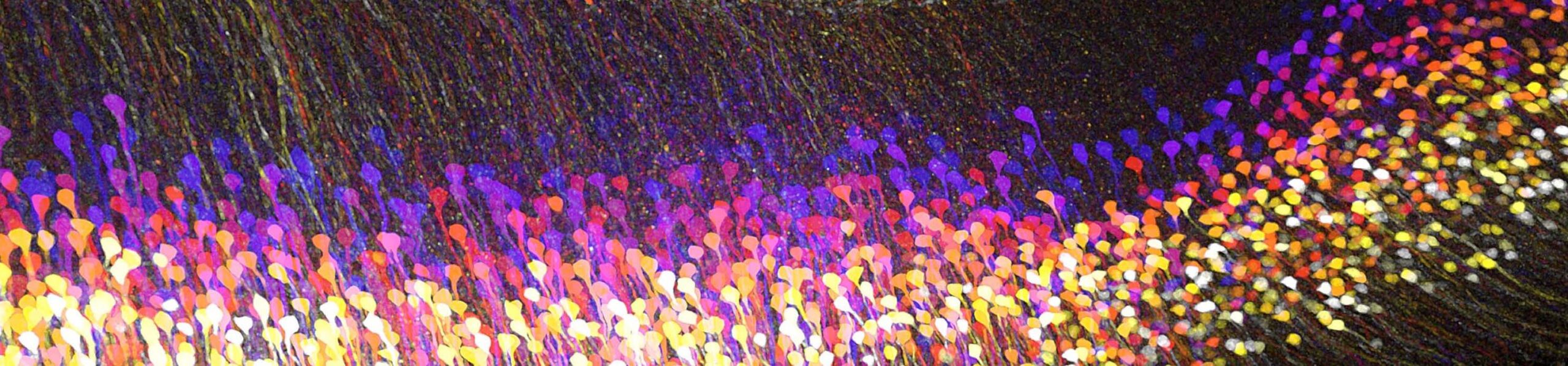
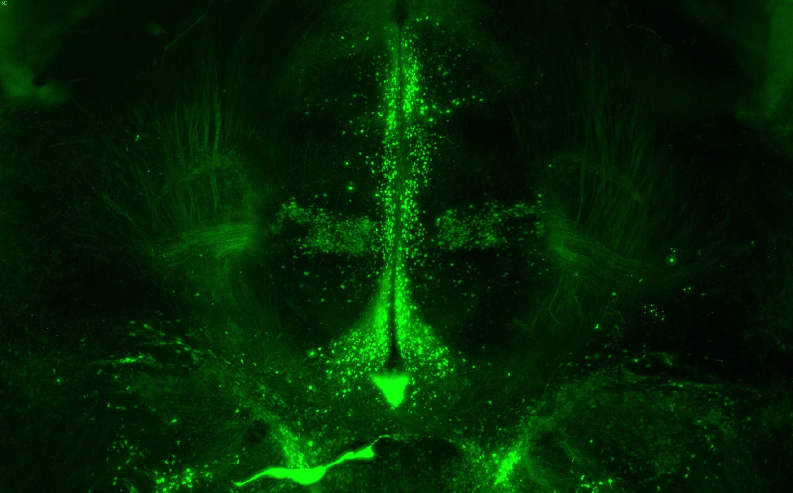
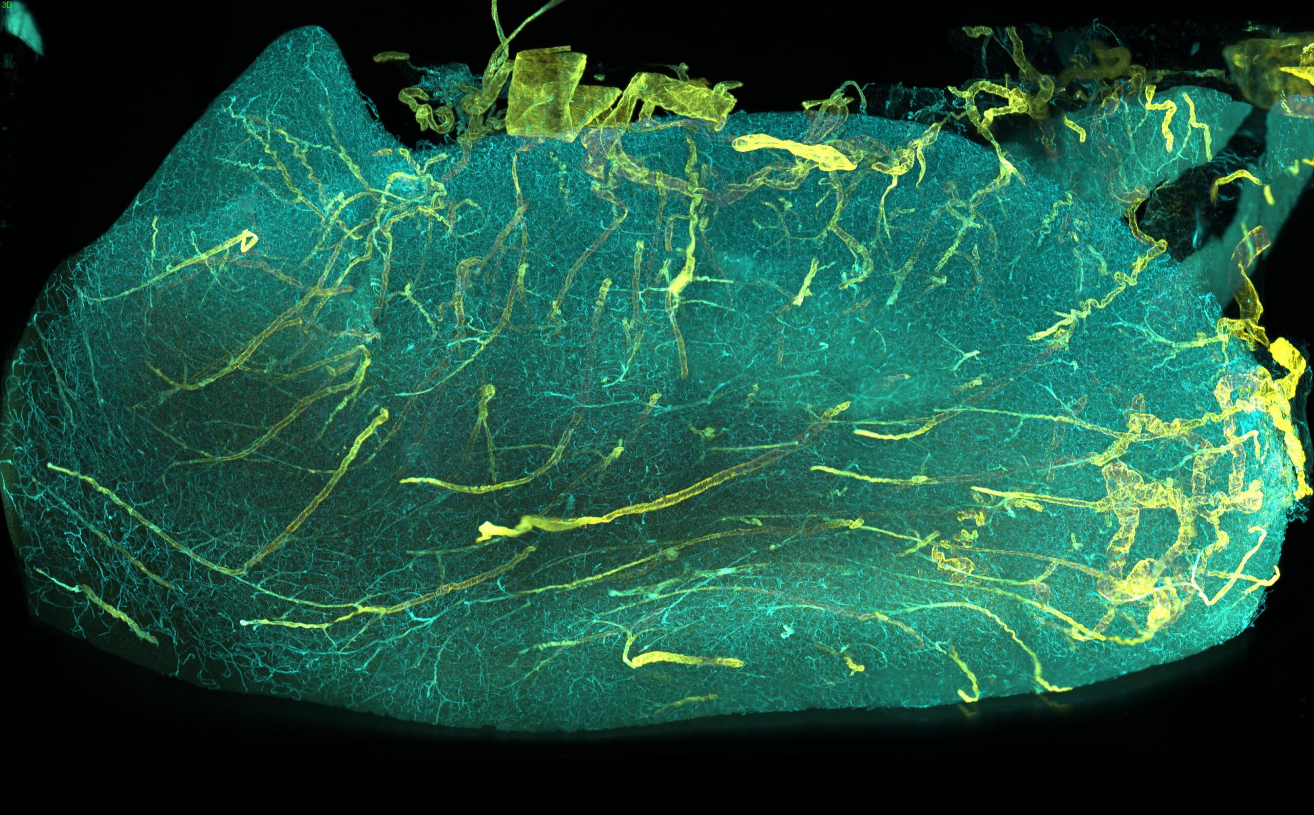
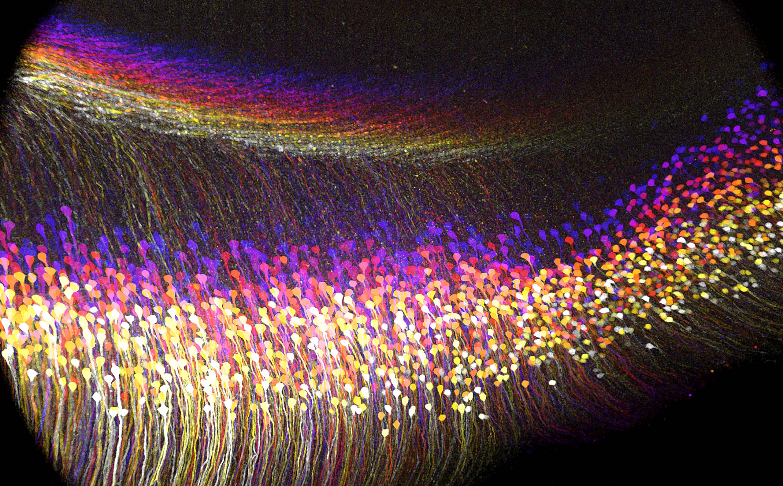
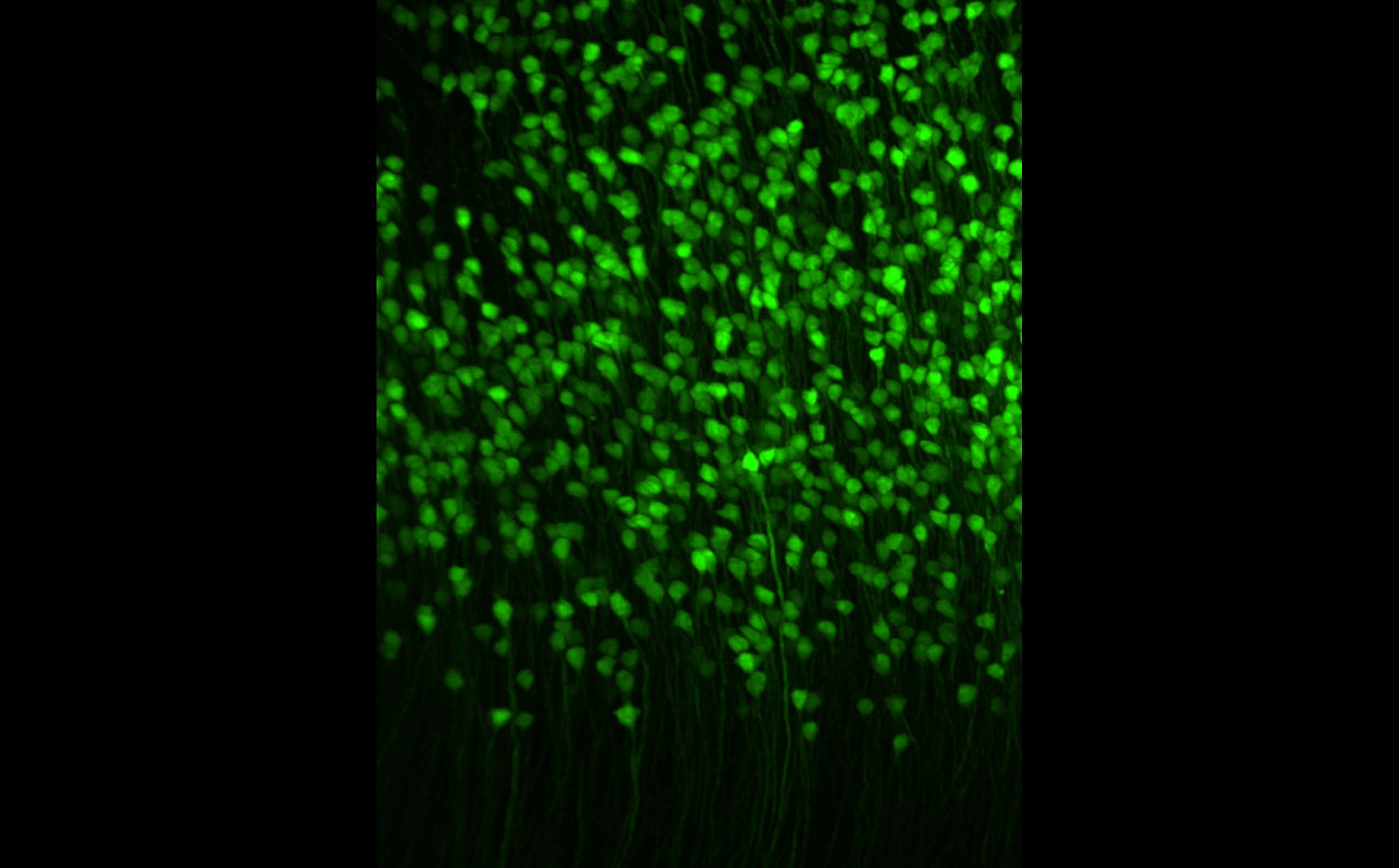
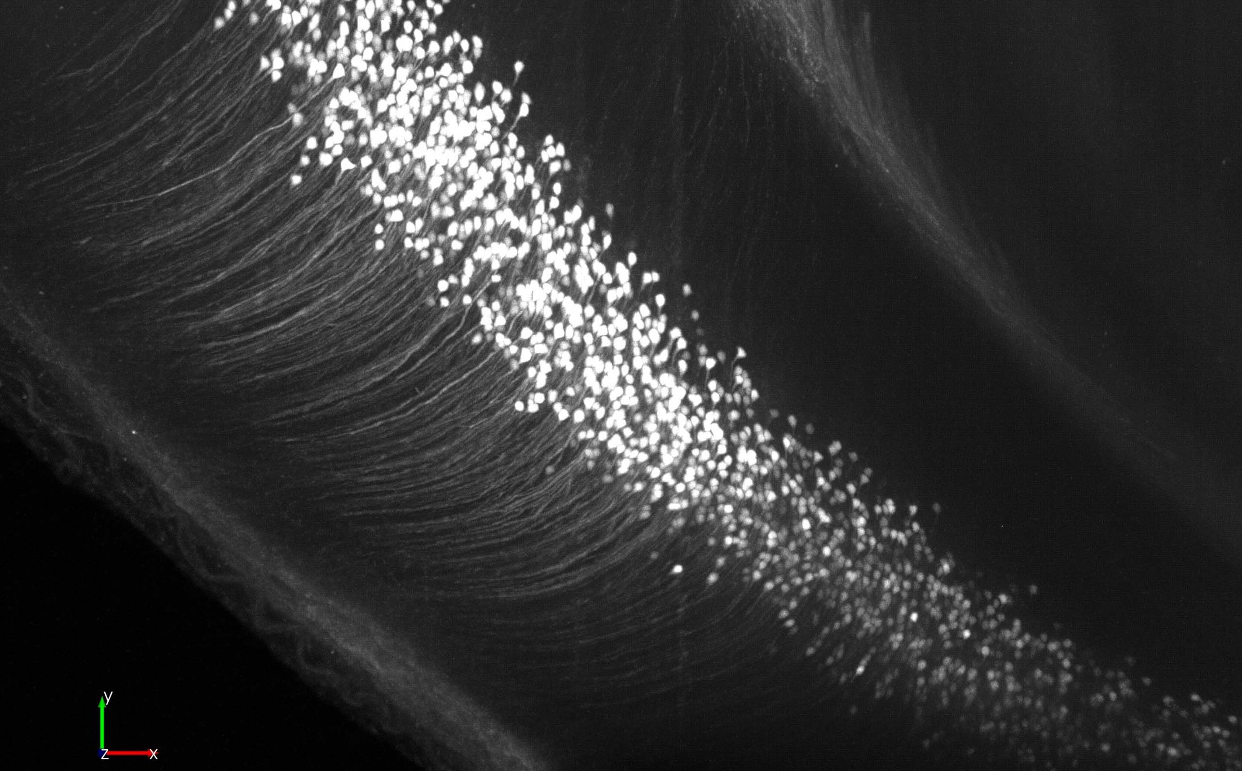
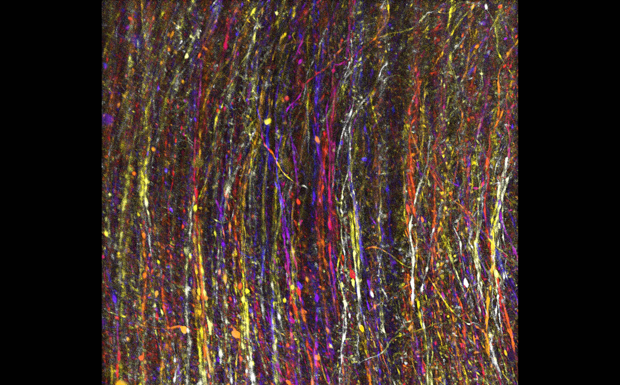
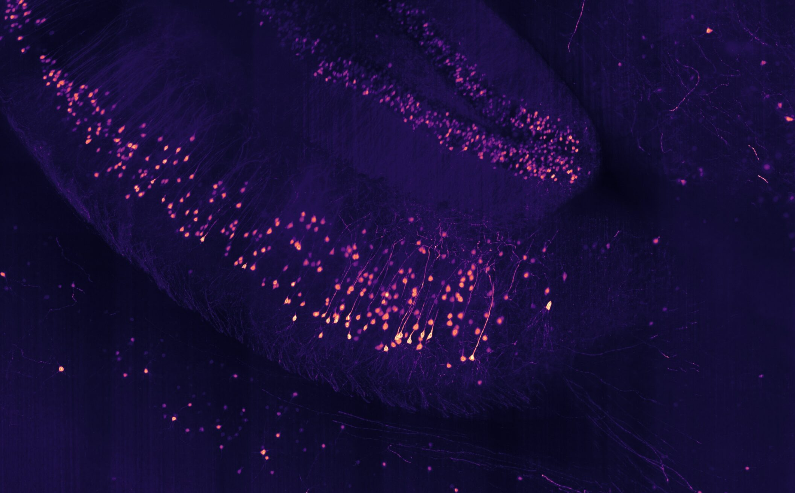
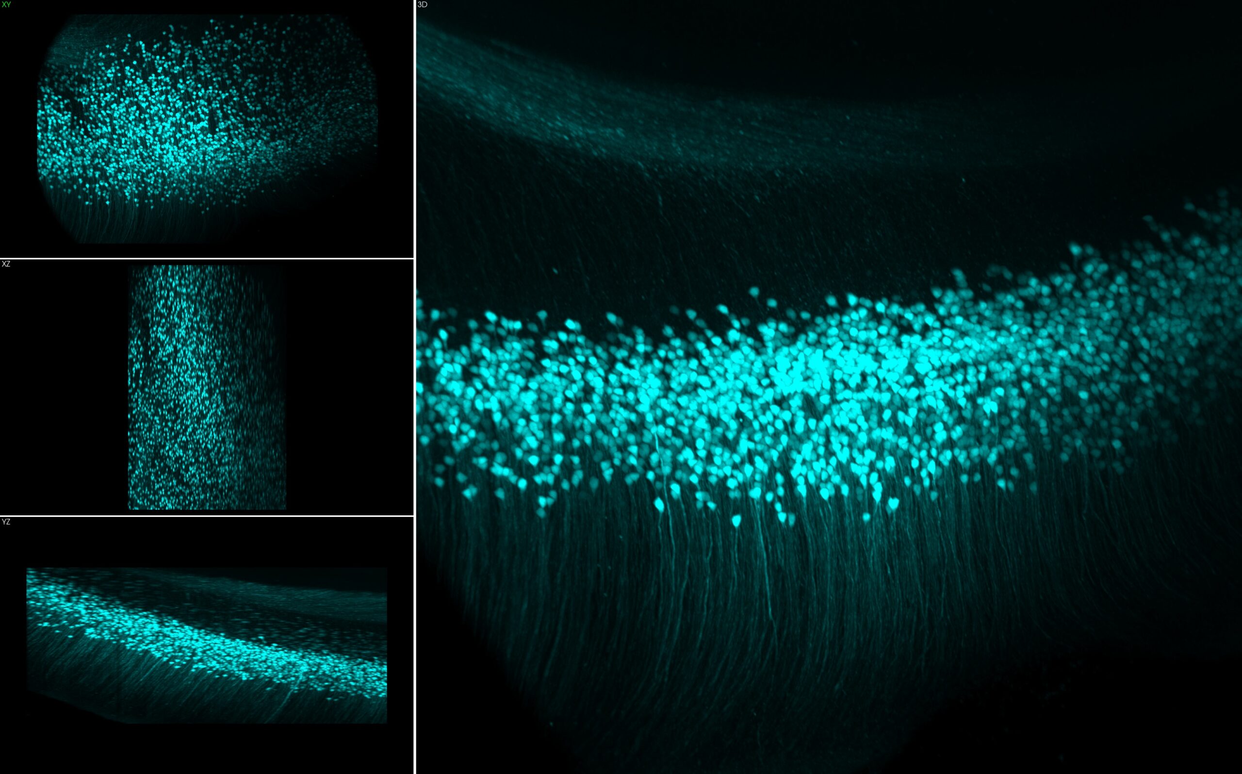
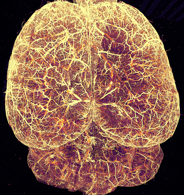
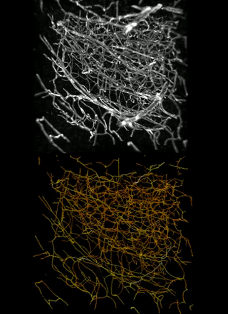
- Image labeled cells and neuronal processes throughout cleared tissue
- 3 illumination wavelengths for popular labels such as: GFP, Thy1-GFP, eYFP, Alexa 514, 633, 647, mCherry, tdTomato
- Compatible with iDISCO, CLARITY, SHANEL, CUBIC, BINAREE and other optical clearing techniques
- Compact design, benchtop ready
Why Choose SLICE?
SLICE offers the power of advanced light-sheet microscopy at a fraction of the cost of conventional systems. Projected Light Sheet Microscopy (pLSM) is a cutting-edge imaging technique utilizing dynamic, modulated light sheets for high resolution visualization of cleared samples. This innovative approach enables multi-resolution imaging, compatibility with a variety of clearing methods and minimal photobleaching making it particularly valuable for fields like developmental biology, neuroscience, and other areas requiring detailed analysis of cleared intact specimens. SLICE embodies these advancements, delivering a compact design and user-friendly interface that makes cutting-edge imaging technology accessible to labs of all sizes.
SLICE delivers unmatched accessibility, giving every team member the freedom to explore, experiment, and uncover new possibilities—without barriers or boundaries. It’s designed to make the extraordinary within reach, offering the ultimate value without sacrificing innovation or quality.
SLICE is engineered so that it can also be placed in locations where experiments are actually happening – e.g. for higher biosafety labels, small lab spaces or even inside incubators.
Versatile Applications
-
-
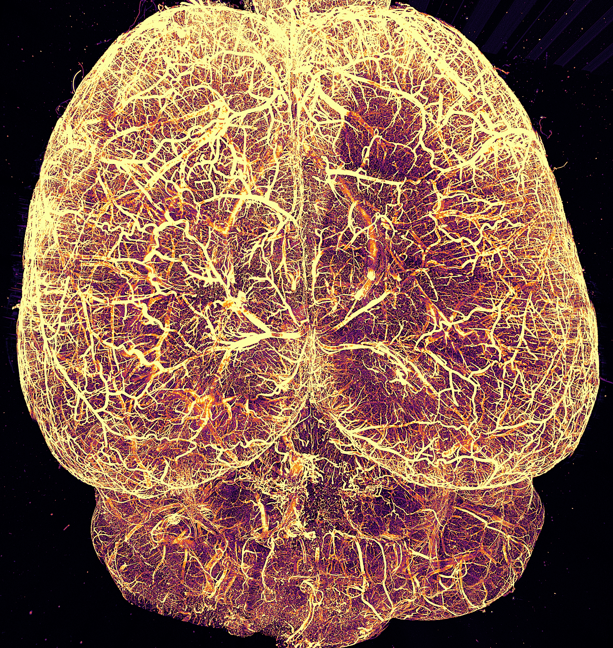
- Whole-brain imaging and mapping
- Vascular system imaging
- Neurodegenerative disease research
- Developmental biology studies
- General biological research requiring 3D imaging
-
Unparalleled Performance
-
-
-
SLICE offers exceptional imaging capabilities:
- Easy to operate and align
- Software-driven light-sheet for fast scan or high-resolution mode
- Minimal photobleaching for extended imaging sessions
-
-
Published, Patent Pending Design
-
-
- Invented by leading microscopy researchers at Columbia University
- SLICE is the next iteration of a peer-reviewed published method, (Chen et al 2024)
- Highlighted in Nature Reviews Bioengineering
-
Advanced Features
-
-
- Linearly Adaptive Light-Sheet Offset Correction: Enables compatibility with a wide array of clearing protocols without the need for any hardware configuration
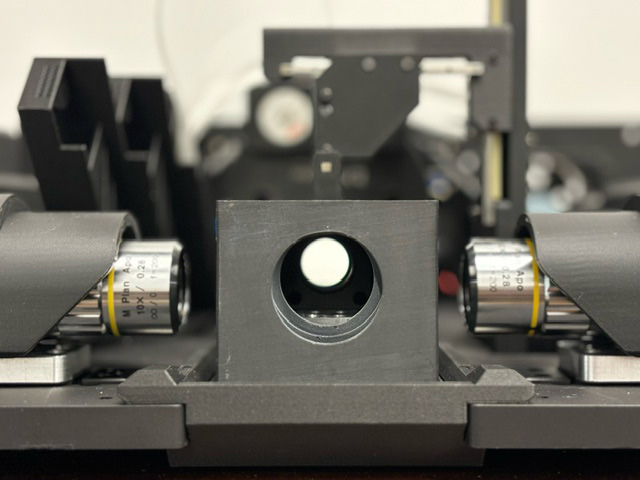
- Multi-View Imaging: Dual opposing light sheets for uniform illumination
- Software-driven multi-resolution imaging
- Automated Multi-Channel Acquisition: Seamless switching between illumination wavelengths
- Software-Driven Light-Sheet Modulation: Adjustable light-sheet thickness from 5 to 50 um for optimized imaging
- Acquisition Software: Streamlines image collection, stitching, post- processing, 3D visualization with add-ons available for atlas registration, neuron quantification, cell counting, deconvolution, etc
- Multi-Size Cuvettes with Sample Holders
- Samples are imaged in exchangeable, fast-mounting, magnetically-held cuvettes designed with users to support the stable imaging of complexly shaped organs and tissues
-
| Lateral Resolution | ~1μm (with 10x objective) |
| Axial Resolution | ~5μm at light sheet waist |
| Light-Sheet Field of View (FOV) | Software controllable; >640 μm |
| Imaging Speed | Up to 40 frames per second* (sample dependent) |
| Multi-Color Imaging | Blue (455nm), Green (520nm), Red (640nm) |
| Physical Dimensions | L 457mm x W 368mm x H 304mm |
| Clearing methods supported | iDISCO, CLARITY, SHANEL, CUBIC, BINAREE and other optical clearing techniques |
| Labeling methods supported | GFP, eGFP, CFP, Thy1-GFP, mCerulean, mTurquoise, eYFP, Citrine, mNeonGreen, Oregon Green, YOYO-1, FITC, SYTOX green and blue, Alexa 514, 633, 647, mCherry, tdTomato, mKate2, Cy5, including c-Fos conjugates, podocalyxin, Acta2, CD31, and others |
| Software-driven multi-resolution imaging |
BrightSlice Software
BrightSlice is a powerful, all-in-one software solution designed specifically for light-sheet microscopy. From acquisition to analysis, handle teravoxel-scale imaging with unprecedented ease and precision.
Core Capabilities
Smart Acquisition
-
- Real-time microscope control with intelligent parameter optimization
- Multi-channel imaging with automated synchronization
- Automated metadata logging for perfect experiment reproducibility
- Save and load custom imaging protocols
Advanced Processing
-
- Automated flatfield correction for uniform illumination
- Seamless stitching of hundreds of image tiles
- GPU-accelerated deconvolution (optional via NeuroDeblur®)
- Sophisticated noise reduction and background correction
- Flexible data compression without quality loss
High-Performance Visualization
-
- Instant viewing of multi-terabyte datasets
- GPU-accelerated 3D rendering
- Real-time maximum intensity projections
- Interactive sub-volume exploration
- Professional-quality movie generation
High-Performance Computing
-
- Highly parallelized architecture
- Advanced GPU optimization
- Efficient memory management
- Smart data streaming
- Optimized memory management
- Industry-standard file formats
- Automated parameter logging
- Comprehensive metadata tracking
Streamlined Workflow
-
-
Everything you need in one intuitive interface:
- Seamless transition from acquisition to analysis
- Batch processing capabilities
- Automated quality control
- Integrated data management
-
Research-Ready Output
-
-
Generate publication-quality data with comprehensive processing tools:
- Multi-resolution data export in JPX and OME-TIFF formats
- Publication-ready visualization
-
Powerful Integration with the MBF Bioscience Ecosystem
-
-
Seamlessly connects with our professional analysis suite:
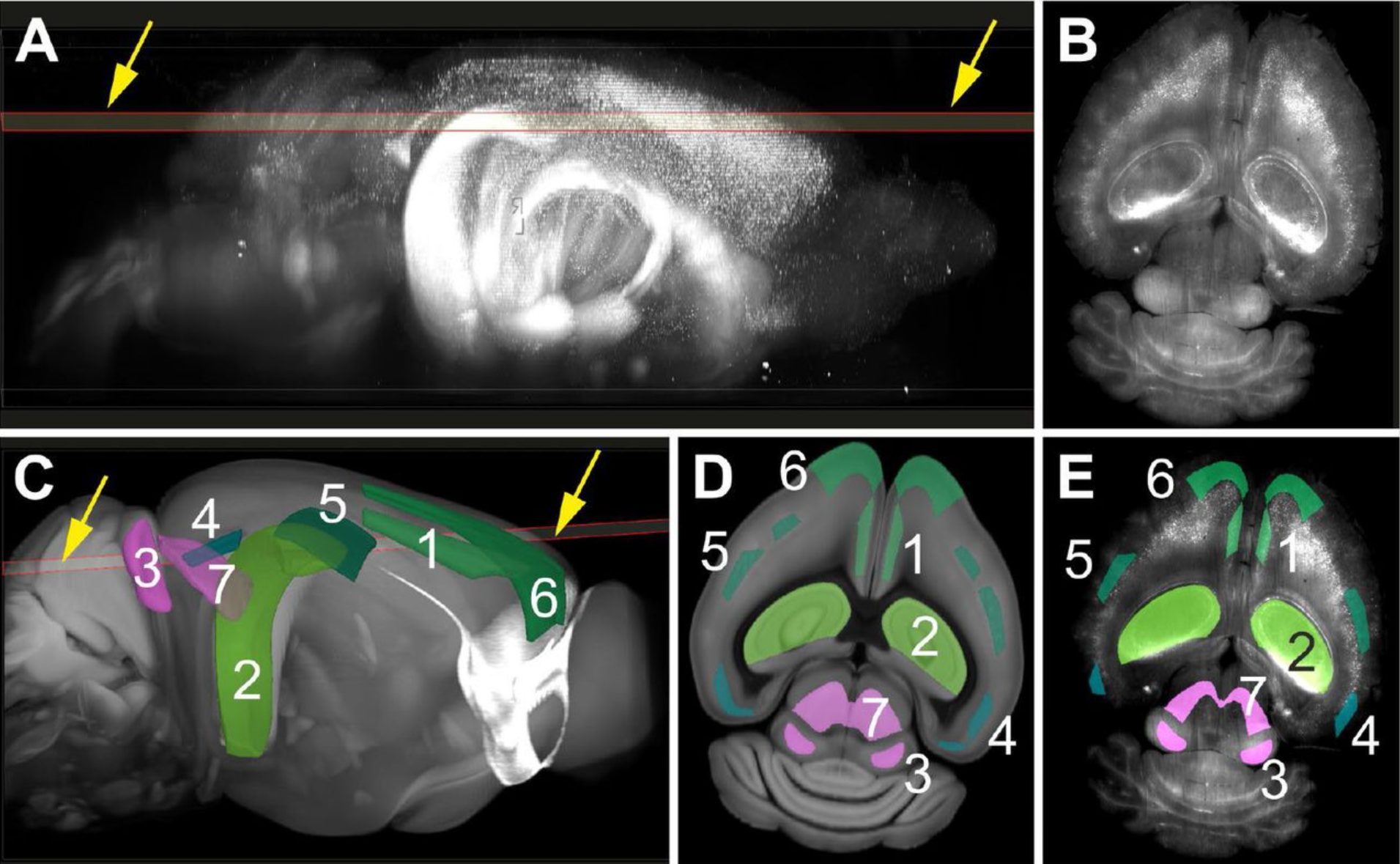
- NeuroInfo®: Advanced brain atlas registration and cell segmentation
- Neurolucida 360®: Industry-leading neuron and vessel analysis
- Stereo Investigator®: Gold-standard stereology
- NeuroDeblur®: State-of-the-art deconvolution
-
Third-Party Compatibility
-
-
Plays well with your existing tools through OME-TIFF export:
- Fiji/ImageJ
- Python
- MATLAB
- Napari
-
Technical Excellence
-
- Parallel processing architecture
- Advanced GPU acceleration
Download SLICE product sheet here.


Sample Preparation and Compatibility
The SLICE system is designed to work with a variety of sample preparation techniques:
- Clearing Methods: Compatible with iDISCO, CLARITY, SHANEL, BINAREE and other optical clearing techniques
- Sample Mounting: Custom-designed oil chamber with RI-matching oil (n = 1.454)
- Multi-Color Imaging: Compatible with commonly used fluorescent proteins and dyes
- Sample Types: Whole mouse brains, human brain sections, organoids, bacterial biofilms
Sample Adaptability
The SLICE offers exceptional flexibility for various imaging needs:
- Adjustable Axial Resolution: from 5 to 50 μm through software-controlled light-sheet thickness
- Scalable Imaging Speed: Trade-off between resolution and speed for different applications
- Multi-Sample Imaging: Optimized for sequential imaging of multiple samples
- Custom Sample Chambers: Adaptable for specific sample types and sizes
BrightSlice Software
Comprehensive Acquisition, Processing and Visualization for Light Sheet Microscopy
SLICE comes with BrightSlice software, a comprehensive software application to handle teravoxel-scale imaging data with ease. From acquisition to visualization, BrightSlice provides everything you need in one powerful, intuitive package:
-
- Light sheet image data acquisition
- Multi-channel imaging
- Post-processing for flatfield correction
- Stitching to create a seamless image compiled from individual image stacks
- Extensive 3D visualization
- Movie creation
- Data compression
Light-sheet fluorescence microscopy generates large image data, which requires powerful acquisition and processing software. BrightSlice software performs all the microscope control and post-processing you will need to create and visualize teravoxel light sheet microscope images. It also offers an optional add-on module for deconvolution.
The BrightSlice software has an easy-to-use interface, combining flexible image acquisition, high-performance 3D data viewing, and powerful post-processing capabilities into a single application with streamlined workflows and batch processing capabilities.
Image Acquisition and Microscope Control
BrightSlice software ensures experiment reproducibility by saving all imaging parameters in image metadata, and configurations can be saved for future experiments. Data output is optimized for MBF Bioscience’s software NeuroInfo (for brain atlas registration), Neurolucida 360 (for neuron and vessel analysis) and Stereo Investigator (for stereology) Output formats also include OME-TIFF to ensure compatibility with common third party image processing software, such as Fiji, Python, Matlab, and Napari.
Smart Acquisition
-
- Real-time microscope control with intelligent parameter optimization and automatic refractive index compensation
- Multi-channel imaging with automated synchronization
- Automated metadata logging for perfect experiment reproducibility
- Save and load custom imaging protocols

High Performance Image Viewing
BrightSlice’s integrated 3D image rich viewing environment allows researchers to inspect the entire dataset directly after acquisition or after processing. The software is highly parallelized and designed to take advantage of advanced GPU graphics cards. This gives users control over their data with key capabilities, including:
-
- Fast viewing of multi-terabyte data sets
- Maximum intensity projections and partial projections
- Highly parallelized for high performance with big image data
- Interactive sub-volume inspection
- Advanced movie generation

Enhanced Imaging Processing and Stitching
BrightSlice software integrates flexible light-sheet microscope control with multi-channel image viewing with and post-processing to achieve the highest quality images. BrightSlice generates high-quality images ready for publications and in-depth exploration by providing the following features:
-
- Stitching hundreds of tiles into a seamless compiled image
- Automated flatfield correction for uniform illumination
- GPU-accelerated deconvolution (optional) for the highest optical clarity
- Sophisticated noise reduction and background correction
- Flexible data compression without quality loss
- Various other processing tools, such as image down-sampling
Seamless Integration
BrightSlice works perfectly with your existing workflow:
MBF Bioscience Light Sheet Suite
-
- NeuroInfo for brain atlas registration and cell segmentation
- Neurolucida 360 for neuron and vessel analysis
- Stereo Investigator for stereology
- NeuroDeblur for deconvolution
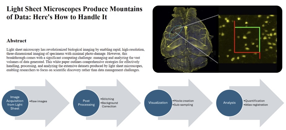
Practical Guidelines for Working with your Large-scale Light Sheet Data
Read our white paper on how to streamline handling, processing, and analyzing extensive datasets from light sheet microscopes, allowing researchers to prioritize scientific discovery over data management challenges.
Ready for Any Challenge
SLICE has been rigorously tested on a wide range of samples:
-
-
- Whole mouse brains
- Human brain tissue samples
- Brain and blood vessel organoids
- Bacterial biofilms
-
Technical Support and Documentation
Our team provides comprehensive support to ensure optimal system performance:
-
-
- Detailed user manual and technical documentation
- Remote installation and setup assistance
- Regular software updates and performance optimizations
- Custom adaptation services for specific research needs
-
Who Is Using MBF Products?
MBF products are used across the globe by the most prestigious laboratories.


Key References
MBF’s products utility is underscored by the number of references it receives in the worlds most important scientific publications. See examples below:
Horejs, CM.
Low-cost, high-performance light-sheet microscopyView Publication
Chen, Y., Chauhan, S., Gong, C. et al.
Low-cost and scalable projected light-sheet microscopy for the high-resolution imaging of cleared tissue and living samplesView Publication
Frequently Asked Questions (FAQ)
What types of labeling can be used with SLICE?
A wide range of proteins and dyes can be used with SLICE, including c-Fos, tdTomato, mCherry, podocalyxin, CD31, Acta2, mScarlet, and Thy1-eYFP. (See the Specifications section.)
What software comes with SLICE?
SLICE comes with BrightSlice software - a full featured application for image acquisition, highly versatile 3D visualization and a rich suite of image optimization tools with state-of-the-art functionalities to support your analysis and publication needs. A quick preview mode enables you to visualize multi-TB image volumes immediately following acquisition. Advanced image stitching produces 3D image volumes suitable for quantitative analysis to produce accurate data for analyzing morphometry, cell populations, sub-cellular processes, vasculature and fluorescence properties.
Will SLICE work with the clearing technique I used for my samples?
SLICE is compatible with a wide array of tissue clearing protocols (e.g. CLARITY, iDISCO, BINAREE, SHANEL, CUBIC, etc.) using intelligent refractive index compensation (IRIC). Users can select the appropriate configuration settings for their tissue sample easily within the intuitive GUI. SLICE uses a linearly adaptive light-sheet offset correction system to correct misalignment between the light sheet and the detection focal plane in large samples, making it compatible with various tissue-clearing methods.
What storage solutions are available for handling the large data files generated by SLICE?
Optimized data storage systems with fast transfer rates take the stress out of working with multi-TB image datasets. MBF Bioscience offers high performance imaging workstations coupled to large capacity, expandable RAID networked attached storage units using 10GB/sec network connections for fast data transfer improving productivity. We can custom configure a network storage solution tailored to your lab's needs.
Why is SLICE so affordable compared to other light sheet microscopes?
SLICE uses a revolutionary, patent pending approach developed by leading researchers at Columbia University, called projected light sheet microscopy. Projected light-sheet microscopy uses digital light projection technology to provide state-of-the-art imaging performance with a compact device footprint, while substantially lowering costs. A recent paper describes this approach: https://rdcu.be/dTCrk
Testimonials
"I rarely have encountered a company so committed to support and troubleshooting as MBF."

Andrew Hardaway, Ph.D. Vanderbuilt University
"MBF Bioscience is extremely responsive to the needs of scientists and is genuinely interested in helping all of us in science do the best job we can."

Sigrid C. Veasey, MD University of Pennsylvania
"I am so happy to be a customer of your company. I always get great help related with your product or not. With the experienced members, you are the best team I've ever met. All of your staff are very kind and helpful. Thank you for your great help and support all the time."

Mazhar Özkan Marmara Üniversitesi Tıp Fakültesi, Turkey
"We’ve been very happy for many years with MBF products and the course of upgrades and improvements. Your service department is outstanding. I have gotten great help from the staff with the software and hardware."

William E. Armstrong, Ph.D. University of Tennessee
"Our experience with the MBF equipment and especially the MBF people has been outstanding. I cannot speak any higher about their professionalism and attention for our needs."
Bogdan A. Stoica, MD University of Maryland
"MBF provides excellent technical support and helps you to find the best technical tools for your research challenges on morphometry."
Wilma Van De Berg, Ph.D. VU University Medical Center - Neuroscience Campus Amsterdam
Robust Professional Support
Our service sets us apart, with a team that includes Ph.D. neuroscientists, experts in microscopy, stereology, neuron reconstruction, and image processing. We’ve also developed a host of additional support services, including:
- Forums
We have over 25 active forums where open discussions take place.
>> Learn More - On-Site/Training
We’ve conducted over 750 remote software installations.
>> Learn More - Webinars
We’ve created over 55 webinars that demonstrate our products & their uses.
>> Learn More
Request more Information
At MBF, we’ve spent decades understanding the needs of researchers and their labs — and have a suite of products and solutions that have been specifically designed for the needs of today’s most important and advanced labs. Our commitment to you is to spend time with you discussing the needs of your lab — so that we can make sure the solutions we provide for you are exactly what you’ll need. It’s part of our commitment to supporting you — before, during, and after you’ve made your decision. We look forward to talking with you!