Celebrating 50 Years of the Computer Microscope (1963-2013) – Part I: The First Semi-Automatic System for Neuroanatomical Analysis
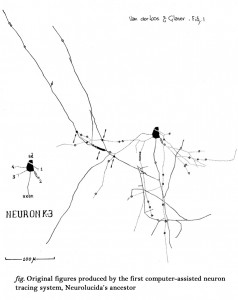
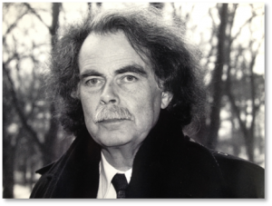
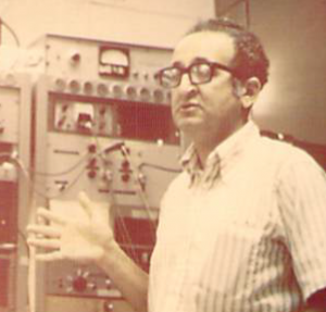
In 1963, Dr. Ed Glaser (co-founder of MBF Bioscience) and Dr. Hendrik van der Loos were at the John Hopkins Medical School putting the final touches on the first computer microscope, an analog computer connected to a light microscope. It was described as a system for attaching X-Y-Z transducers to a microscope stage, tracing the branches of a Golgi-stained neuron and outputting the result to a plotter. The microscope stage was equipped with high-linearity linear motion transducers yielding output voltages proportional to the position of the microscope stage in all three dimensions. The computation of distances was performed by using a chord approximation to the curvilinear dendrites whose lengths were to be determined.
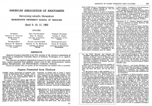
In April of 1963, at the annual meeting of the American Association of Anatomists at the Georgetown University School of Medicine, Drs. van der Loos and Glaser presented a paper, “Semi-automatic quantitation of dendrite characteristics in the cerebral cortex,” in which they unveiled their “specially designed semi-automatic apparatus” that could “be considered of use, in general, in these investigations in which three-dimensional measurements of microscopic distances is required.”
The computer microscope, invented to run van der Loos and Glaser’s software for neuron tracing and analysis (Neurolucida’s ancestor), would drastically improve neuronal morphometric studies, as we will explain in our next post.


