Neurolucida®360

The most advanced software for automatic 3D neuron tracing and reconstruction
MBF Bioscience > Neurolucida®360

Product Overview

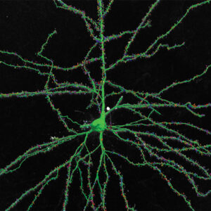
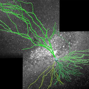
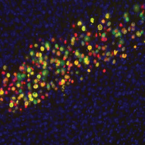
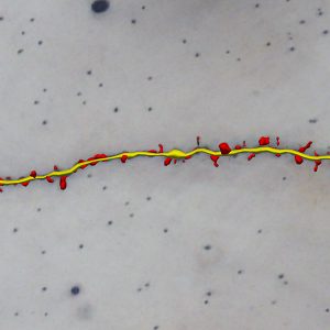
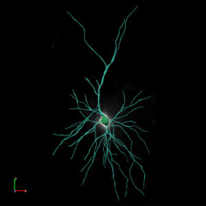
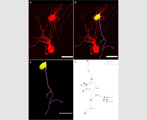
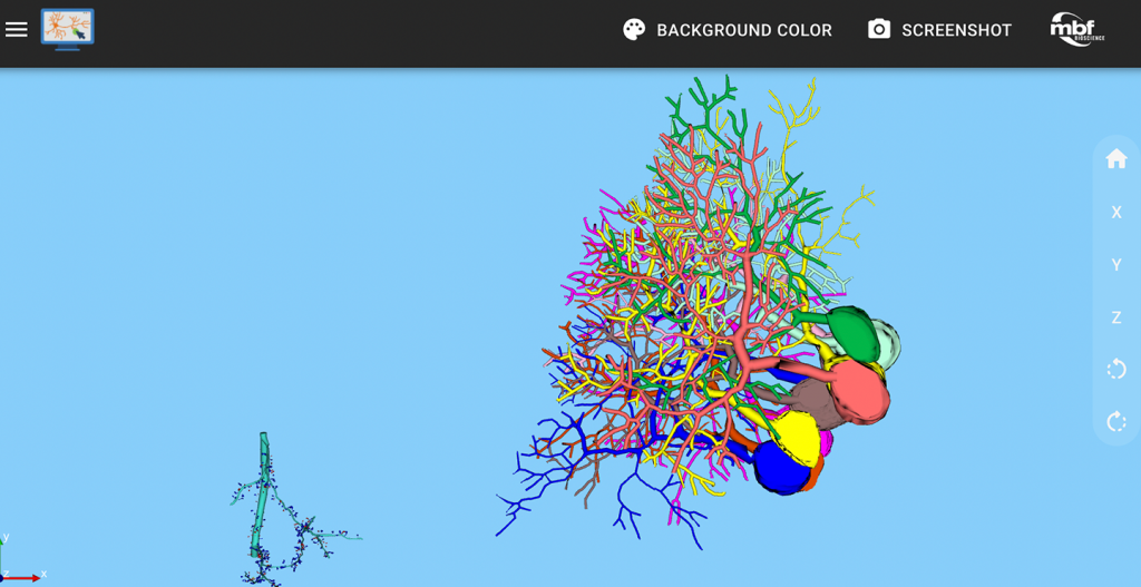
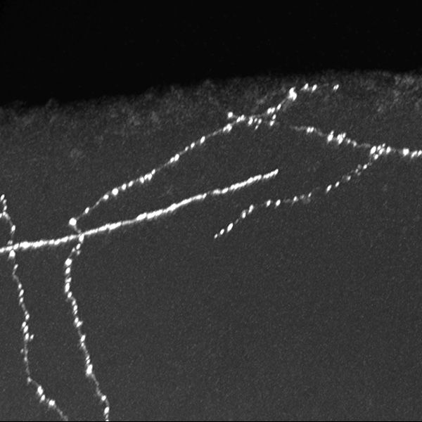
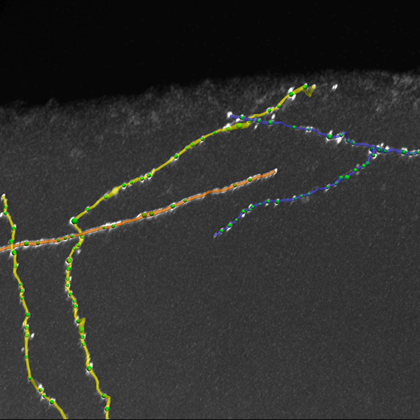
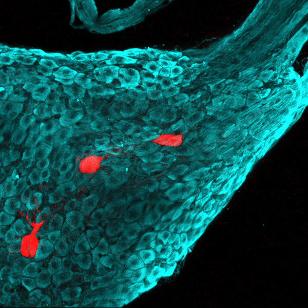
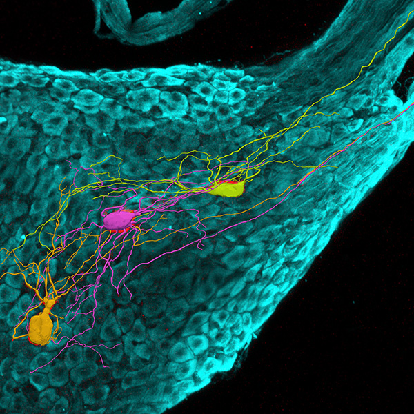
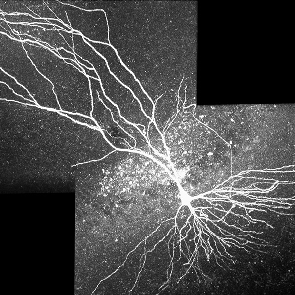
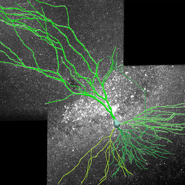
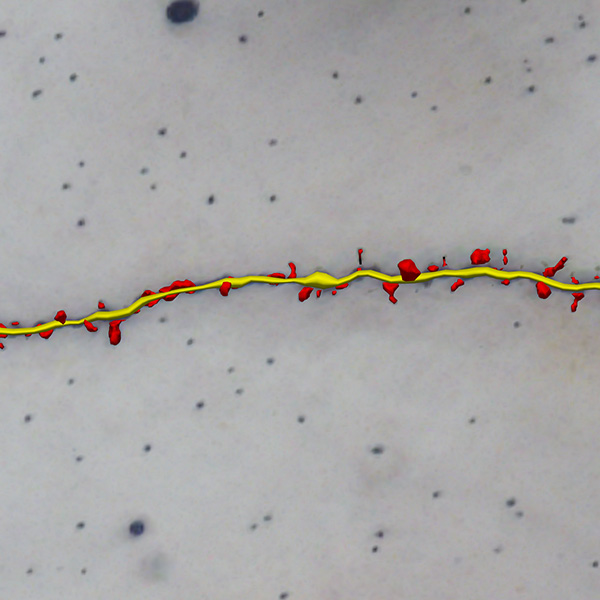
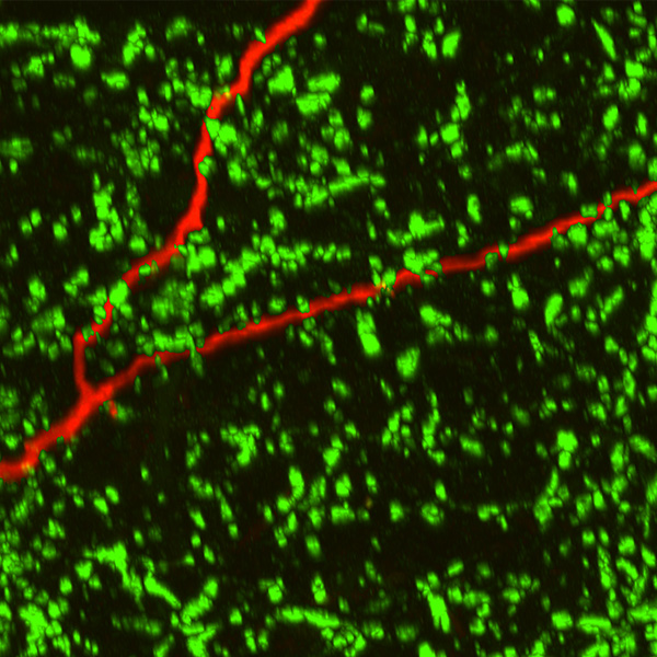
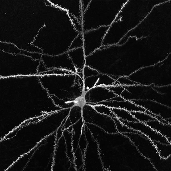
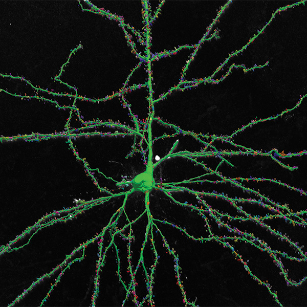
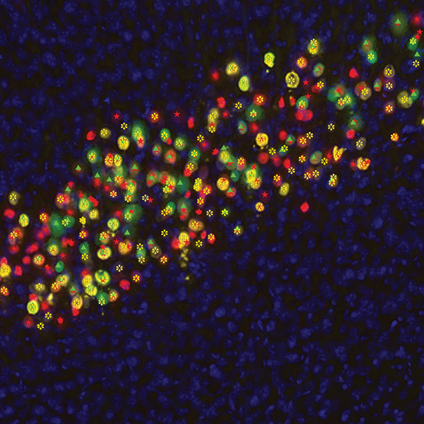
Neurolucida 360 is the premier software used by neuroscientists to quickly and accurately reconstruct and trace any neuron from any species using a wide variety of labeling and microscopy techniques. With the most advanced 3D image detection algorithms coupled with machine learning techniques, Neurolucida 360 performs accurate reconstruction of neuronal structures that range in scale from complex, multicellular neuron networks, to sub-cellular neuronal components such as dendritic spines, varicosities and putative synapses. This comprehensive software also includes methods to analyze glial cells such as oligodendrocytes, microglia, and astrocytes.


Neurolucida 360 is engineered to work efficiently with even the largest 3D multi-channel image datasets using our innovative intelligent precognitive image handling technology. And, it is compatible with file formats acquired from most research microscopes.

Product image NL360

Key Benefits

Neurolucida 360: The leading image analysis software for automatic 3D neuron tracing and reconstruction and quantitative morphology

Neurolucida 360 is the most trusted software for comprehensive neuron tracing and reconstruction. Built by neuroscientists for neuroscientists, Neurolucida 360 is equipped with powerful automated algorithms for detection of neuronal structures present in 2D and 3D microscopy image data.

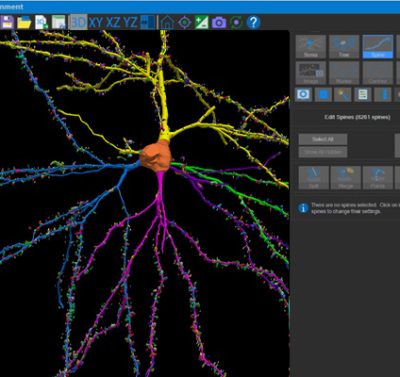
With built-in workflows, an intuitive user-interface, and interactive 3D environment, Neurolucida 360 makes it easy to reconstruct somas, axons, dendrites, varicosities, spines, and synapses.

Paired with the companion software, Neurolucida Explorer, you can quickly and effectively analyze the morphology of subcellular structures, whole cells, or complex connective networks – from any species.

Neurolucida 360 supports many types of histological preparation techniques and microscopy modalities, such as:

  • Immunofluorescence
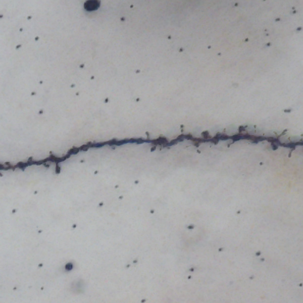
  • Golgi-Cox
  • AAVs
  • Retrograde or anterograde filled cells
  • Immunohistochemistry
  • Brightfield
  • Widefield fluorescence
  • Confocal
  • Light sheet
  • 2-Photon, Multi-Photon
  • Whole brain or organ
  • Cleared tissue
  • Sectioned tissue
  • Cell cultures
  • Expanded tissue


Neurolucida 360 has been developed with support from the National Institute of Mental Health (NIMH)

Ultra is here! A 3D automatic vessel tracing module for thorough exploration of neuro-vascular environments.

Recommended Hardware Requirements

Neurolucida 360 is a powerful software that can work with a wide range of image data of vastly different levels of size and complexity. Because of its flexibility, our recommendations for system configurations vary to balance the affordability of lower computing power for smaller data sets to more expensive high-end workstations for large, multi-channel data.

Please contact us to consult with our technical specialists for a more precise recommendation specific to your data needs.

64-bit Windows 11 operating system
CPU with 8 cores (16 threads) or more. More cores improve performance when using large data sets (>1 GB).
Solid state hard drive(s). Preferably, non-volatile memory express (NVMe) drives.
64 GB of system memory or more. More memory is better for large data sets, especially for image handling and reconstruction in the 3D environment.
Graphics card with 8 GB memory or more. Most graphics cards from NVIDIA and AMD have been tested with MBF Bioscience software.
Minimum Hardware Requirements
64-bit Windows 10 operating system
8 core processor (16 threads)
32 GB memory
Graphics card with 6 GB memory or more: This is the minimum needed for the 3D environment, but is adequate for working only in 2D.
Computer-Hardware Upgrade Priorities
To upgrade your system for better performance with MBF Bioscience software, we suggest that you prioritize computer hardware upgrades as follows:
      • Increase system memory to at least 128 GB. More memory improves performance when using large data sets
      • Replace hard disk drives (HDDs) with solid-state drives (SSDs), preferably non-volatile memory express (NVMe) drives
      • If you're working with 3D images and graphics, upgrade graphics card (GPU) to one with at least 8-12 GB memory

From Proteins to Dendritic Spines: Neurolucida 360 Plays a Crucial Role in Advancing Neuroscience
>> Learn More

Blue Brain scientists develop pyramidal cell classification system using neurons reconstructed with Neurolucida 360
>> Learn More

Curcumin Lowers Neuroinflammation in Mouse Model
>> Learn More

Researchers Identify Potential Treatment for Patients at Risk for Alzheimer’s Disease
>> Learn More

MBF Bioscience research team contributes novel dendritic spine analysis in study published in Science
>> Learn More

NeuroMorpho.Org Releases Nearly 10,000 New Neuron Reconstructions and Neurolucida leads the way
>> Learn More


Visit our blog to read more case studies

Download Neurolucida 360 product sheet here.

Version 2024.2.1 (September 2024)

New Features and Enhancements

  • New Puncta-detection user interface improves and expands capabilities for specifying puncta detection settings:
    • Detect puncta based on proximity to other structures, such as trees, spines, and varicosities using rule sets
    • Employ powerful And/Or criteria in your rule sets to find specific puncta easily
    • Save your settings and reuse your puncta-detection criteria for multiple images, including in the batch pipeline for processing entire directories of image data
  • Measure Intensity export will now report the centroid coordinate of any puncta
  • You can now enable or disable the detection of spines and varicosities in the same location
  • New ability to detect somas in the same location in different color channels
  • The typical process width setting is now available in the automatic tracing setup
  • New image filters added to the batch process pipeline:
    • ExM image filter includes three model options to use to optimize modeling of your expansion-microscopy images
    • Tone Mapping image filter to reveal details in both bright and dim regions of your image
  • The 3D Environment has an added option to eliminate the display of edge artifacts at the surface of 3D volumes
  • Automatic stitching of overlapping fields of view in the Montage tool is now available to optimize the visual information of compiled image montages
  • Improvements to the image support in the Measure Intensity tool
  • Image support for the .czi format has been improved
  • Slide scans from MBF Bioscience acquisition software can now be compiled
  • Improvements to the movie creation tool:
    • You can now generate movies at high resolution for large data sets
    • Use the image slice and partial projection views during movies to showcase important information
    • Simple drag and drop to open .xml movie files


See a full list of new features here 

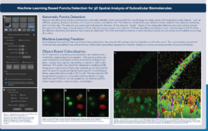
This is a Neurolucida 360 poster

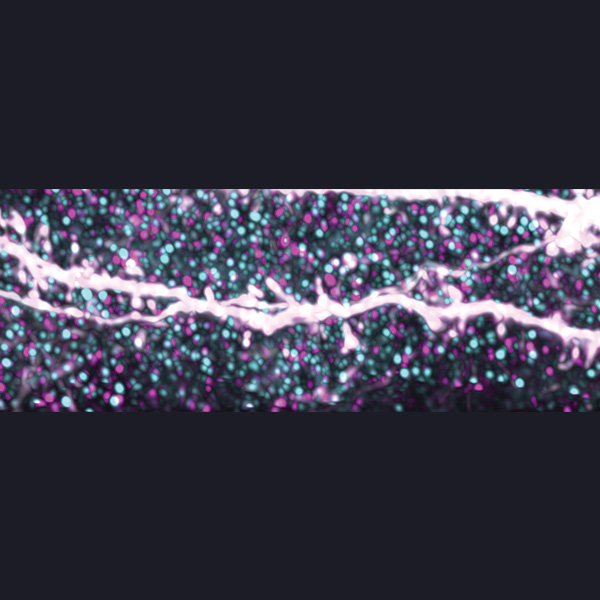
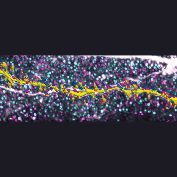
An AI-Extended Solution for Performing Integrated 3D Neuro Microstructure and Nanostructure Analysis of Expansion Microscopy Data

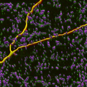
Automatically reconstruct whole cells, dendritic spines, puncta, and varicosities with Neurolucida 360

< >
< >
< >
< >
< >
< >
< >
< >
< >

Introducing: Neurolucida 360 Ultra!​

Empowering Comprehensive Exploration of Neuronal and Vascular Interactions 


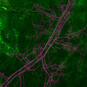
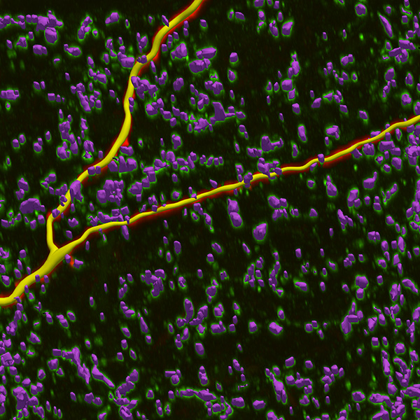
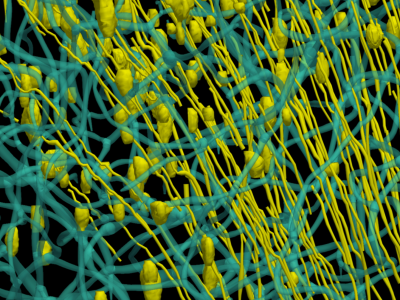
We are thrilled to unveil Neurolucida 360 Ultra software. Now, you can segment, reconstruct, and analyze blood vessels and microvasculature – in addition to neuronal structures. By integrating the advanced vascular analysis capabilities from Vesselucida 360 into Neurolucida 360, the new Neurolucida 360 Ultra package provides you with a single, advanced application that offers an unparalleled opportunity to explore the intricate dynamics of neuronal and vascular structures.

Vessels&Neurons-smaller

Affordable Packages

Our three most popular packages are Neurolucida 360 Lite, Essentials, and Studio.

There are different options to set up Neurolucida 360 in your lab: a one-time upfront cost for the software, an annual subscription plan, mobile licenses to move Neurolucida 360 from one computer to another, and more.

Feature Description

Neurolucida 360
LITE

Neurolucida 360 ESSENTIALS

Neurolucida 360 STUDIO

Neurolucida 360
ULTRA

User-guided tree tracing
Smart manual tree tracing
User-guided soma detection
Set specified color channels for analyses
3D editing
Create and share movies
Professional technical support
Open and FAIR file format
Big image data capability: Batch image filters
Big image data capability: Sub-volume tool
Big image data capability: Dense area visualization tool
Big image data capability: Dense area automatic tracing
Automatic tree tracing
Batch mode
Automatic soma detection
Image montaging
Automatic puncta detection, including synapses
Automatic puncta detection, with machine learning
Automatic spine detection and classification
Automatic trace evaluation
Automatic vessel detection

Who Is Using Neurolucida 360?

Neurolucida 360 is used across the globe by the most prestigious laboratories.

News About Neurolucida 360

Cited in Peer Reviewed Scientific Publications

Neuroluicda 360’s utility is underscored by the number of references it receives in the worlds most important scientific publications. See examples below: 

Frequently Asked Questions (FAQ)

How affordable is Neurolucida 360?

To fit your scientific and budgetary needs, we have three popular packages of Neurolucida 360 available for purchase: Lite, Essentials, and Studio.

View comparison chart

There are even different options to set up any of the above Neurolucida 360 packages in your lab, including:  annual subscription plans, mobile licenses to move Neurolucida 360 from one computer to another, and more.

What microscopy file formats are supported by Neurolucida 360?

Our software supports almost all microscopy image file formats generated from a variety of microscopy vendors (CZI, TIF, JPX, LIF, and more). See the comprehensive list here.

Want to convert your file format to standardized formats, JPEG2000 (.jpx/.jp2) and/or OME-TIFF? Try our free tool, MicroFile+, which uses state-of-the-art compression methods to efficiently and accurately convert 2D and 3D images from most sources into manageable and metadata-enriched format(s).

How can I quantify the neuronal tracings and reconstructions created in Neurolucida 360?

With the included companion analysis Neurolucida Explorer software, you can perform detailed morphometric analyses of your Neurolucida 360 reconstructions. Automatically obtain quantitative data from a plethora of morphological, spatial (e.g. Sholl Analysis), and orientation analyses. In addition to the sophisticated and comprehensive generation of quantitative data, Neurolucida Explorer also shows many graphical displays that allow you to visualize the quantitative data in intuitive ways – and generate figures for publications and presentations.


What’s the difference between Neurolucida 360 and Neurolucida?

Neurolucida 360 can automatically reconstruct neurons from images and image stacks acquired by a variety of microscopy types – whereas Neurolucida – Microscope Edition enables manual reconstruction of neurons from tissue specimens directly from a research microscope.


I have hundreds of images to use with Neurolucida 360 – can I process data in batches?

Yes! Our Neurolucida 360 Studio package has sophisticated batch tracing tools for detecting somas, trees, varicosities, spines, and puncta across multiple files.

To further expedite your data throughput, you can also apply image filters in batch and employ batch analysis features with Neurolucida Explorer.

Are there tools in the software for creating high quality movies and images for publications?

Yes, you can create dynamic, high-resolution MP4 videos of your tracing and image data in Neurolucida 360’s intuitive 3D environment.

Are there online resources to help me learn the software?

All MBF Bioscience software comes with comprehensive, context-sensitive help guides accessible online or offline. Further, reduce the initial learning curve with our online learning center for how-to videos, quick guide PDFs, and product-specific webinars.

How should I prepare and image neurons for analysis with Neurolucida 360?

Neurolucida 360 excels at handling multi-resolution image data from a wide range of tissue preparations (e.g., serial sectioned, cleared), microscopy modalities (e.g., confocal, light sheet, expansion microscopy). Depending on your reconstruction and analysis goals, our team of experts can help you refine imaging parameters for optimal performance in Neurolucida 360. Contact us today.

Additionally, our scientists collaborated with researchers from the Icahn School of Medicine at Mount Sinai in New York to develop a set of guidelines on preparing and imaging confocal image data for Neurolucida 360. The guidelines, published in Current Protocols in Neuroscience, will help you get the best results for spine quantification and neuron reconstruction.

+ Read the publication

Testimonials

Robust Professional Support

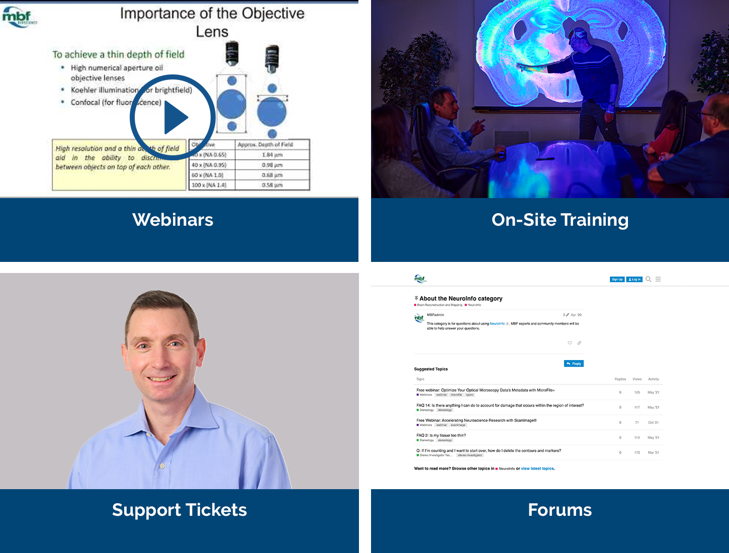
Our service sets us apart, with a team that includes Ph.D. neuroscientists, experts in microscopy, stereology, neuron tracing and reconstruction, and image processing.  We’ve also developed a host of additional support services, including:

  • Forums
    We have over 25 active forums where open discussions take place.
    >> Learn More
  • On-Site/Training
    We’ve conducted over 750 remote software installations.
    >> Learn More
  • Webinars
    We’ve created over 55 webinars that demonstrate our products & their uses.
    >> Learn More

Request an Expert Demonstration

We offer both a free expert demonstration and a free trial copy of Neurolucida 360. During your demonstration you’ll have the opportunity to discuss your hardware, software, or experimental design questions with our team of Ph.D. neuroscientists and experts in microscopy, neuron tracing, and image processing.


During your free trial, use the tips and suggestions from a free, expert evaluation and find out how easy Neurolucida 360 is to use and how quickly you can obtain useful data.

Related Products

Neurolucida®

Neuron tracing & analysis directly at the microscope. The gold standard for neuron tracing.


Neurolucida® Explorer

The analytical software companion for Neurolucida and Neurolucida 360 designed to perform extensive morphometric analysis.

Vesselucida® 360

Automatically reconstruct microvascular networks in a 3D environment.

ClearScope® - The Light Sheet Theta Microscope

Ground-breaking light sheet microscope system for cleared specimens.

Recorded Webinars and Videos

Introduction to Neurolucida 360

Webinar: Reconstructing Neuron and Vessel Morphology in Cleared Tissue

Webinar: Introducing Neurolucida 360’s Enhanced Batch Pipeline